New stem cell serum extract promises wrinkle reduction in weeks – full sized product worth £176 to be won

A new and potent anti-aging breakthrough derived from non-embryonic human stem cell research has just been launched in Europe. And Elixir has a samples of this exclusive Blue Horizon Stem Cell Serum to give away in our competition. All you have to do is answer a simple question at the end of this article.

Blue Horizon promises a 23% reduction in fine lines, wrinkles and scars in just two weeks. Although the serum does not contain actual human stem cells, its formula is based on stem cell research. The company offers stem cell treatments outside of the US in unique collaborations with scientists.

The main and unique active ingredient in the serum are stem-cell derived proteins called cytokines which send signals to regulate cells and regenerate skin.  It also contains proven skin anti-ageing ingredients including epidermal growth factors which are natural combination of skin growth promoters; short and long-chained hyaluronic acid and ceramides to combat the effects of aging and deliver unique skin benefits without surgery.

The serum has been toxicology tested within the EU and independent research on twenty individuals aged 46 to 81 found a 23% reduction in skin roughness, including a decrease in the appearance of fine lines, wrinkles and scars in just two weeks. The test was carried out by Dermatest®, a top, independent German pharmaceutical and cosmetic test accreditation laboratory, who used Dermatological Optical 3D Test:Norm DIN 4768 on the skin of 20 test subjects using the serum. The serum is made in Germany.

If you want to get your hands on a pot of this unique anti-ageing serum you can here
at the Blue Horizons website Although its not cheap at £176 ($295
) for 30 ml which is approximately one-month’s supply, the cream has been flying off the shelf.

If you would like to win a full size sample please answer a simple question in our competition below:

click here to find out more at the Blue Horizons website

Patients own skin may be used to repair damage of Mutiple Sclerosis

Scientists have revealed that a patient’s own skin may be able to repair the nerve damage caused by the incurable disease, Multiple Sclerosis.
MS attacks the fatty lining around the nerves causing malfunctions of movements and more.
But scientists at the University of Rochester Medical Centre in the US have used skin cells to repair the damage in  animal tests, says a report in the journal Stem Cell.
Just like electrical wires, nerves have insulation – but instead of plastic, the body uses a protein called myelin.
However, diseases that result in damage to the myelin, including MS, leave the nerves exposed and electrical signals struggle to travel round the body.
They took a sample of skin cells and converted it into stem cells, which are capable of becoming any other type of cell in the body. The next step was to transform the stem cells into immature versions of cells in the brain that produce myelin.
When these cells had been injected into mice born without any myelin it had had a significant effect, said researchers.
However, MS patients would still have the problem of their immune system continuing to attack their myelin.
Any treatment would need to be used alongside other therapies to tame the immune system – or would need to be repeatedly performed.
Enhanced by Zemanta

Scientists harvest living stem cells from dead

Scientists have harvested stem cells from the scalps and brain linings of human corpses and reprogrammed them into stem cells.
Stems.jpeg
Their discovery – that  dead people can yield living cells that can be converted into any cell or tissue in the body, has been recognised with the Nobel Prize.
The researchers believe that this discovery could lead to new stem cell therapies for the treatment of mental disorders, such as schizophrenia, autism and bipolar disorder, which may stem from problems with development.
Mature cells can be made or induced to become immature cells, known as pluripotent stem cells, which have the ability to become any tissue in the body and potentially can replace cells destroyed by disease or injury. 
Cadaver-collected stem cells are collected from fibroblasts (skin cells) and can be reprogrammed into induced pluripotent stem cells using chemicals known as growth factors that are linked with stem cell activity. 
Reprogrammed cells could then develop into a multitude of cell types, including the neurons found in the brain and spinal cord. 
Now scientists have taken fibroblasts from the scalps and the brain linings of 146 human brain donors and grown induced pluripotent stem cells from them as well.
Researcher Thomas Hyde, a neuroscientist, neurologist and chief operating officer at the Lieber Institute for Brain Development in Baltimore said that they were now able to culture living cells from the deceased  on a far larger scale than previously.
The bodies had been dead up to nearly two days before scientists collected tissues from them. The corpses had been kept cool in the morgue, but not frozen. The researchers found fibroblasts taken from the brain lining, or dura mater, were 16 times more likely to grow successfully than those from the scalp. This was expected, since the scalp is prone to fungal and bacterial contamination just like any other part of the skin. 
These contaminants can ruin any attempt to grow fibroblasts in lab dishes.
Surprisingly, scalp cells did proliferate more and grew more rapidly than dura mater cells. 
“This makes sense — the skin is constantly renewing, while the turnover in dura mater is much slower,” Dr Hyde said.
Future therapies
Cells from corpses might play a key role in developing future stem cell therapies. Successfully reprogramming induced pluripotent stem cells so they behave like the cells they are meant to replace means that samples of the mimicked cells must be present for comparison. Cadavers can provide brain, heart and other tissues for study that researchers cannot safely obtain from living people.
“For instance, we can compare neurons derived from fibroblasts with actual neurons from the same individual,” Hyde said. “It tells us about how reliable a given method for deriving neurons from fibroblasts is. That can be crucial if, for example, you want to create dopamine-making neurons to treat someone with Parkinson’s disease.”
Studying how induced pluripotent stem cells develop into various tissues could also shed light on disorders that are due to malfunctions in development.
“We’re very interested in major neuropsychiatric disorders such as schizophrenia, bipolar disease, autism and mental retardation,” Hyde said. “By understanding what goes wrong with the brain cells in these individuals, we could perhaps help fix that.”
Enhanced by Zemanta

Scientists create functioning human livers

New York: Researchers have created miniature replicas of human livers which functioned normally in laboratory conditions. 

The development may  eventually solve the transplant shortage and also remove the need for powerful drugs to prevent the body rejecting the organ. But it will be at least five years before the technology can be used in hospitals.

The project director, Associate Professor Shay Somer of the Wake Forest Institute for Regenerative Medicine in North Carolina said: “We are excited about the possibilities this research represents, but must stress that we’re at an early stage and many technical hurdles must be overcome before it could benefit patients.

“Not only must we learn how to grow billions of liver cells at one time in order to engineer livers large enough for patients, but we must determine whether these organs are safe to use in patients.”

it is estimated that more than a fifth of patients die waiting for a transplant and many livers have to be discarded because they are too old or too damaged to be of any use.

The technology opens up the prospect of growing other replacement organs, including kidneys or pancreases, for patients who are able to donate stem cells.

Pedro Baptista, co-author, said: “Our hope is that once these organs are transplanted, they will maintain and gain function as they continue to develop.”

The new technique works by effectively chemically stripping the old liver down too its basic “scaffold” or exoskeleton in a process of called “decellularisation”.

Onto this frame of connective tissue and blood vessels, they then regrow the new liver using stem cells from the patient.

Laboratory livers that were nourished for a week began growing and functioning like human organs, they said.

Liver disease is the fifth biggest killer in England and Wales, after heart disease, cancer, stroke and respiratory disease, and the only major cause of death that is still increasing year on year.

Some 16,087 people in Britain died from liver disease in 2008, a 4.5 per cent increase on the previous year, and the number of deaths is predicted to double in 20 years.

Sarah Matthews, for the British Liver Trust, said: “Technology such as this is much needed. Currently supply isn’t meeting demand, and for every one person who receives a liver transplant, 10 people die.

liverpetri.jpgThe research was presented at the annual meeting of the American Association for the Study of Liver Diseases in Boston.


Gene cocktail creates beating heart cells

Los Angeles: A new technique, in which a gene cocktail, has been successfully to create beating heart cells, has been used on mice.

The new technique could also cut the need for transplants in people whose ailing hearts cannot mend themselves.

heart.bmpIf developed for use in human patients the new treatment could change the lives of many thousands of sick people.

The experiments, which were carried out on mice, are still in the early stages, but they offer fresh hope for the future.

They centre on the large muscular cells that allow the heart to beat and go about its vital work of pumping blood. Normally, the body has little or no way of replacing any that die or are damaged.

But researchers at the California’s Gladstone Institute of Cardiovascular Disease, have devised a cocktail of genes that trick other heart cells called fibroblasts into transforming into beating muscle cells called cardiomyocytes.

The journal Cell reports how fibroblasts treated with the cocktail in the lab turned into beating muscle cells after being transplanted into a mouse.

Masaki Ieda of the Gladstone Institute said: “Scientists have tried for 20 years to convert non-muscle cells into heart muscle. It turns out we just needed the right combination of genes in the right dose.”

Deepak Srivastava, senior study author from Gladstone, said: “The ability to re-programme fibroblasts into cardiomyocytes has many therapeutic implications.

“Half of the cells in the heart are fibroblasts, so the ability to call upon this reservoir of cells already in the organ to become beating heart cells has tremendous promise for cardiac regeneration.”

In time a drug that works in the same way as the cocktail of genes could be developed. Injected into damaged hearts, it would drive the growth of new muscle.

“That’s our long-term goal,” said Srivastava.

Every two minutes, someone, somewhere has a heart attack. Many go on to develop heart failure, in which the weakened heart gradually loses its ability to pump blood.

Up to 40 per cent of these die within a year of diagnosis – giving heart failure a worse survival rate than many cancers.

Other organs could be patched up in a similar way, the researchers believe.


Man’s eyesight restored with his own stem cells

image

London: A 38-year-old-man has had his vision restored with stem cells after being blinded in an ammonia attack.

Russell Turnbull, 38, lost the sight in one eye in 1994 when he was squirted with ammonia after trying to stop a row on a bus in Newcastle.

He was left with Limbal Stem Cell Deficiency (LSCD), a painful condition which requires constant treatment.

The new treatment was developed by scientists at Newcastle University.Their method involves taking a small amount of stem cells from a patient’s good eye, cultivating them in a laboratory, and implanting them into the damaged cornea.

He was was treated at Newcastle’s Royal Victoria Infirmary (RVI) and is one of of eight patients who successfully underwent the treatment developed at the North East England Stem Cell Institute (NESCI).

It is hoped that the technique could eventually be rolled out into clinics.

Scientists create jaw bone from stem cells

image

New York: Scientists have created part of the jaw bone for the first time in the lab using human adult stem cells.

The technique, which created the joint, may be useful for other areas of the body.

The Columbia University study appears in Proceedings of the National Academy of Sciences.

The bone which has been created in a lab, by scientists at Columbia University is called the temporomandibular joint (TMJ). This joint which is complex to repair can be damaged by defects at birth, arthritis or injury. Treatment is notoriously difficult as the joint has a complex structure and is difficult to replicate using bone grafts.

Scientists used human stem cells taken from bone marrow which were then planted into a “tissue scaffold” in the shape of a human jaw bone.

The cells were then cultured nutrients found during natural bone development.

Lead researcher Dr Gordana Vunjak-Novakovic said: “The availability of personalised bone grafts engineered from the patient’s own stem cells would revolutionise the way we currently treat these defects.”

She said the new technique could also be applied to other bones in the head and neck, including skull bones and cheek bones, which are similarly difficult to graft.

She stressed that the joint created in the lab was bone only, and did not include other tissue, such as cartilage which the team is also working on.

Italian surgeon unveils stem cell facelift pictures

image
image

Dr. Renato Calabria, pioneer of the Stem-Cell Enhanced Facelift has unveiled before and after pictures of the procedure he performed in Florence, Italy earlier this Spring. While it’s still early, the results will only get better over time!

Here Dr Calabria explains the procedure:

For decades plastic surgeons have been looking to restore a more youthful look on the face. Surgical techniques have been improving, newer lasers have been used in order to achieve a goal that seemed unreachable. Comparing the post-operative results with the pictures of the patients when they were younger, it appeared that there was something missing: a younger face had more volume and the skin just looked fresher.

Dr. Calabria always thought that with the advent of stem cell research, plastic surgery could beneficiate from it. Using stem cell as a mean to jump start the regenerative process in our own body was definitely a very attractive proposition.

We knew that the fat in our body contained adult stem cell along with other regenerative cells. Dr. Calabria has been using fat grafting ( therefore containing adult stem cells) on the face with the distinct purpose to rejuvenate the skin and adding volume.

But was not till the develop of a new device, the Celution Device, that facial rejuvenation could taken to the next level and finally create a really amazing result.

Dr.Calabria has called this new way of the future the “cell-enhanced face lift”.

The (stem) cell-enhanced face lift is a new procedure used to volumizing the face and create a more youthful look.

Beverly Hills Plastic Surgeon Dr. Renato Calabria believes that traditional face lift should be a thing of the past: they caused the so called “wind tunnel “look by pulling tight the skin in the wrong direction. Adding volume to the face has always been Dr. Calabria’s goal. First with the Vertical Face lift, by moving the patient’s own tissues in a vertical direction and therefore repositioning it to the original and more natural position, Dr Calabria has been creating a more youthful look. And now with the (stem) cell-enhanced face lift, Dr. Calabria think he has definitely found the answer.

The (stem) Cell-Enhanced face lift not only restores the volume in the face but also adds regenerative cells which are believed to be beneficial to the rejuvenation process.

Dr. Calabria has developed a technique by which the cells are injected into the SMAS and underneath the facial musculature to create a more youthful volume adding result.

The (stem) cell-enhanced face lift combines soft tissue that is transferred from one part of the body to the face with the patient’s own adipose tissue-derived stem cells and regenerative cells.

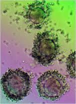
The technique first consists of harvesting the cells ( Usually from the lower abdomen). Liposuctioned fat is rich with regenerative cells which include adult stem cells, blood vessel producing cells, growth factor secreting cells. Once the fat is harvested, it is treated through a device to isolate the cells.

Celution® Technology – How it Works
First, adipose tissue is taken from the patient by a low volume tissue collection procedure. Next, the collected tissue is placed in the Celution® Device, which processes each patient’s tissue with a single-use, application-specific consumable set that easily attaches to the Celution® Device.

The Celution® Device liberates the stem and regenerative cells from the adipose matrix; the cells are then separated, washed, and concentrated in the collection container. This real-time processing takes place in a closed environment to minimize the risk of exposure to contaminants and can be completed within the timeframe of a single surgical procedure.

Next, a syringe is used to collect the cells that are to be redelivered to the same patient. The cells may be injected directly into a site or implanted with a delivery matrix or scaffold to improve performance, such as combining the cells with an adipose tissue graft in reconstructive surgery procedures.

Following re-infusion into the body, it is thought that environmental cues from the damaged and surrounding tissue guide the stem and regenerative cells to the area of damage and help facilitate a natural healing response. The cells may respond by a variety of mechanisms, which promote tissue survival, graft retention, and/or differentiation of progenitor cells into the appropriate cell type.

Then, during the face lift, the regenerative cells tissue embedded in fat is then gently placed with a blunt cannula in layers of facial tissues like the SMAS, under the muscles, etc…. Once the fat is placed, symmetry is checked and then the skin is redraped and approximated, the excess skin excised and then anchored in a vertical direction and closed in layers. The key is to place this regenerative tissue in a precise location in order to enhance the volume of the face in areas previously volume deprived.

After the cell-enhanced face lift, the patient exhibits a dramatic improvement not only in the underlying soft tissue contouring of the face but the skin itself. The growth factors contained in these cells induce the skin and the other tissues to produce more cells of their own by basically initiating a signal to the local stem cells as well as the transplanted adult stem cells to restore them self and multiply.

Others techniques of stem cell face lift inject the cells directly in the face,( without surgery) but in Dr. Calabria’s opinion is not as effective in voluminizing the face that the cell-enhanced ( stem cell) face lift and it does not address the laxity of the skin.

Also the cells have not been isolated with the celution device which Dr. Calabria thinks is essential to the final result.

The regenerative cells, by secreting growth hormone and other factors, are also beneficial to create a more youthful look, diminishing the aging process. The procedure restores the youthful contour and shape of the face as well as skin tightness and evens out color irregularities caused by aging and sun damage.

The cell-enhanced (stem cell) face lift is the new frontier of face lifting and Dr. Calabria is definitely writing a new chapter in facial rejuvenation.

The Celution device is not available in the US yet but has been approved for clinical use in Europe and it is available in Dr. Calabria’s Milan and Rome locations and he will bring the technology to the U.S. as soon as next year.

Find more information on Dr Calabria’s website www.drcalabria.com

US scientists use stem cells to reverse MS

image

Chicago: US researchers have reversed multiple sclerosis symptoms in early stage patients by using bone marrow stem cell transplants to reset the immune system.

Commenting on the study, Helen Yates, Chief Executive of the UK’s Multiple Sclerosis Resource Centre said: “This further piece of research into the use of stem cells with Multiple Sclerosis patients provides another piece of evidence that stem cells could one day provide clear therapies and treatments for MS. MSRC hopes that further work in this area proves as positive as this piece of research”

Some 81 percent of patients in the early phase study showed signs of improvement with the treatment, which used chemotherapy to destroy the immune system, and injections of the patient’s bone marrow cells taken beforehand to rebuild it.

“We just start over with new cells from the stem cells,” said Dr. Richard Burt of Northwestern University in Chicago, whose study appears in the journal Lancet Neurology.

Multiple sclerosis occurs when the immune system mistakenly attacks the myelin sheath protecting nerve cells. It affects 2.5 million people globally and can cause mild illness in some people and permanent disability in others.

Symptoms may include numbness or weakness in the limbs, loss of vision and an unsteady gait.

“MS usually occurs in adults,” Burt said in a telephone interview. Before they get the disease, their immune systems work well, he said, but something happens to make the immune system attack itself.

His approach is aimed at turning back the clock to a time before the immune system began attacking itself.

Burt said the approach — called autologous non-myeloablative hematopoietic stem-cell transplantation — is a bit gentler than the therapy used in cancer patients because rather than destroying the entire bone marrow, it attacks just the immune system component of the marrow, making it less toxic.

Burt and colleagues tried the treatment on 21 patients aged 20 to 53 with relapsing-remitting multiple sclerosis, an earlier stage in the disease in which symptoms come and go.

Patients in the study were not helped by at least six months of standard treatment with interferon beta.

After an average follow-up of about three years, 17 patients improved by at least one measure on a disability scale, and the disease stabilized in all patients.

Patients continued to improve for up to 24 months after the transplant procedure, and then stabilized. Many had improvements in walking, vision, incontinence and limb strength.

“To date, all therapies for MS have been designed and approved because they slowed the rate of neurological decline. None of them has ever reversed neurological dysfunction, which is what this has done,” Burt said.

Other teams have seen improvements in patients using a more aggressive approach. In one study led by Dr. Mark Freedman of the University of Ottawa last year, 17 MS patients treated with the more aggressive approach were showing signs of remission two years after treatment.

Burt stressed that the treatment approach needed to be tested in a more scientifically rigorous randomized clinical trial, in which half of the patients get the transplant treatment and the other half get standard treatment.

That trial is under way.

Find more information at www.msrc.co.uk

Stem cells used to repair breast post cancer ops

image

London: British surgeons are using stem-cell-enriched fat from women’s bodies to repair the damage of surgery following breast cancer operations and radiotherapy.

The procedure, which is being trialled at Leeds General Infirmary, so far appears to restore the softness and suppleness of breast tissues, as well as the painful aftermath of treatments.

Each year around 31,000 British women undergo operations to remove cancerous tissue which normally leaves an unsightly cavity in the breast. Some surgeons have already used fat transfer to repair the damage as well as reducing the size of the other breast to match the damaged one.

Scientists believe that fat enriched with stem cells reduces inflammation and helps maintain a healthy blood supply so that the body’s repair system works more efficiently.

Fears that the stem-cells might encourage more cancer cell proliferation have also proved groundless.

The cancer patient’s own fat cells are harvested and made into a concentrate which is reinjected. More than 90% of the fat survives the process.

Lead investigator and consultant plastic surgeon Eva Weiler-Mithoff says she is impressed with the results so far. “What is striking is the softness and suppleness the technique gives the skin and tissues. When I see these stem-cell-enhanced patients after three months, their skin is significantly softer.”

Stem cell hope for MS sufferers

image

Chicago: Stem cell injections may be able to help reverse the crippling effects of multiple sclerosis, a study published today says.

Four out of five adults in the early stages of MS who were injected with stem cells taken from their bone marrow saw an improvement in symptoms after three years, while the condition of the remainer stabalised.

MS is one of the most common disabling neurological conditions,and caused by damage to the myelin – a protective sheath surrounding nerve fibres and results in problems with sensation and muscle control.

The study, at the Feinberg School of Medicine, was designed to see whether injections of stem cells from bone marrow would migrate to parts of the nervous system damaged by MS and repair them.

Among the 21 men and women in the trial,who were aged between 20 and 53, 17 had improved on a scale of disability after three years. None of them reported a worse score.
The report in The Lancet Neurology medical journal today says the technique suppresses cells that cause damage and effectively ‘resets’ the immune system.
Study leader Dr Richard Burt of Feinberg School of Medicine, Chicago, said: ‘It is a feasible procedure that not only seems to prevent neurological progression, but also appears to reverse neurological disability.’
And a further trial involving 100 patients is to get under way soon.

Scientists reverse brain defects in animal models

image

Tel Aviv: Brain birth defects have been successfully reversed, using stem cells, in animal models by scientists at the Hebrew University of Jerusalem.

Neural and behavioral birth defects, such as learning disabilities, are particularly difficult to treat, compared to defects with known cause factors such as Parkinson’s or Alzheimer’s disease, because the prenatal teratogen – the substances that cause the abnormalities — act diffusely in the fetal brain, resulting in multiple defects.

Prof. Joseph Yanai and his team at the Hebrew university-Hadassah Medical School were able to overcome this obstacle in laboratory tests with mice by using mouse embryonic neural stem cells. These cells migrate in the brain, search for the deficiency that caused the defect, and then differentiate into becoming the cells needed to repair the damage.

The stem cells may develop into any type of cell in the body, however at a certain point they begin to commit to a general function, such as neural stem cells, destined to play a role in the brain/ nervous system. At more advanced developmental stages, the neural stem cells take on an even more specific role as neural or glial (supporting) cells within the brain/ nervous system.

In the researchers’ animal model, they were able to reverse learning deficits in the offspring of pregnant mice who were exposed to organophosphate (a pesticide) and heroin. This was done by direct neural stem cell transplantation into the brains of the offspring. The recovery was almost one hundred percent, as proved in behavioral tests in which the treated animals improved to normal behavior and learning scores after the transplantation. On the molecular level, brain chemistry of the treated animals was also restored to normal.

The researchers went one step further. Puzzled by the stem cells’ ability to work even in those cases where most of them died out in the host brain, the scientists went on to discover that the neural stem cells succeed before they die in inducing the host brain itself to produce large number of stem cells which repair the damage. This discovery, finally settling a major question in stem cell research, evoked great interest and was published earlier this year in one of the leading journals in the field, Molecular Psychiatry.

The scientists are now in the midst of developing procedures for the least invasive method for administering the neural stem cells, which is probably via blood vessels, thus making the therapy practical and clinically feasible.

Normally, stem cells are derived from individuals genetically different from the patient to be transplanted, and therefore the efficacy of the treatment suffers from immunological rejection. For this reason, another important avenue of the ongoing study, toward the same goals, will be to eliminate the immunological rejection of the transplant, which will become possible by taking cells from the patient’s own body — from a place where they are easily obtained — by manipulating them to return to their stem cell phase of development, and then transplanting them into the patient’s brain via the blood stream. One important advantage of this approach will be to eliminate the controversial ethical issues involved in the use of embryo stem cells.

The research on the project has been supported by the US National Institutes of Health, the US-Israel Binational Science Foundation and the Israel anti-drug authorities.

Stem stell advance voted scientific discovery of 2008

image

London: A new advance in harvesting stem cells from adults which has the potential to cure many of the diseases of ageing, has been hailed as the scientific discovery of the year.

The advance, which involves turning back the clock on adult tissue and “reprogramming” it with the properties of stem cells, could lead to new treatments for diseases including Parkinson’s and diabetes.

The process allows for a potentially limitless numbers of “induced pluripotent stem” (IPS) cells to be made to order from a sick patient’s cells, meaning they do not risk rejection from the immune system when transplanted.

The technique does not require stem calls to be harvested from embryos, making it more acceptable to religious groups.

Dr Robert Coontz of the journal Science, which placed cellular reprogramming top of its list of the biggest scientific breakthroughs of 2008, said it “opened a new field of biology almost overnight and holds out hope of life-saving medical advances”.

Three teams working in Japan and the United States made major advances with the technique over the last 12 months.

“When Science’s writers and editors set out to pick this year’s biggest advances, we looked for research that answers major questions about how the universe works and that paves the way for future discoveries,” Dr Coontz said.

Runner-up was the first direct observation of planets in distant star systems, which required complex measures to blot out the light from their parent stars.

Other advances on the list included improved technology to map the genome – the human genetic code – and new calculations of the weight of the world.

Science’s Top 10 breakthroughs of 2008

1) Cellular reprogramming

2) Observation of planets around stars

3) Insights into “good” fat

4) Expanding the catalogue of cancer genes

5) Most detailed video of a developing embryo

6) Faster, cheaper genome sequencing

7) Watching proteins at work

8) Industrial-scale energy storage

9) High-temperature superconductors

10) Calculating the weight of the world

Stem cells used to grow frog eye

image

New york: In an experiment at the SUNY Upstate Medical University, in Syracuse, NY researchers have grown frog eyes from stem cells.

Researcher Michael Zuber and his colleagues took the cells from frog eggs.Then got them to become eyes, by genetically modifying them, inserting transcription factors (proteins that trigger expression of other genes) which are known to regulate eye growth and development.

The scientists then implanted the cells into tadpoles missing an eye. The cells properly developed and differentiated into all seven types of retinal cells and appeared to have the proper structure. Additionally the new eye attached properly to the brain. In swimming tests the eye was shown to be working as implanted tadpoles only swam to the white side of the tank (normal behavior), while blind ones would also swim to the black side of the tank.

Would the technique work on mammals? The answer is maybe — frogs naturally have a much easier type regrowing tissues than humans, in fact they can be triggered to regrow legs and many amphibians can regrow lost tails. Triggering proper differentiation in mammals is much more complex.

Nonetheless, Professor Zuber hopes that chemicals will be found from the research that can activate transcription factors in humans. Even if a full eye could not be grown, this could help people with retinal disorders regenerate ocular tissue.

In a separate, but perhaps equally intriguing study performed by Sujeong Jang of Chonnam National University, in South Korea, and his colleagues, the researchers were able to restore the hearing of deaf guinea pigs by implanting them with human neural stem cells obtained from human bone marrow.

Scientists grow windpipe to renew a woman’s life

image

London: Scientists have used stem cells to grow part of a windpipe which was later implanted into a woman whose own trachea had been destroyed by tuberculosis.

The breakthrough procedure, which happened in Barcelona, Spain, is described in online in the British medical journal, The Lancet.

Claudia Castillo, 30, a mother of two living in Barcelona had been suffering from tuberculosis for years. The disease destroyed part of her trachea, the windpipe connected to the lungs. In March, her left lung collapsed and Castillo needed regular hospital visits to clear her airways which left her unable to take care of her children.

Doctors had planned to remove her entire left lung but instead, Dr. Paolo Macchiarini, head of thoracic surgery at Barcelona’s Hospital Clinic, proposed a windpipe transplant instead. He was the one performing the surgery on Castillo.

With the help of a new technique developed at the University of Padua, Italy, scientists removed all the cells from the trachea of a 51-year old donor by essentially scrubbing it clean with a high-tech detergent solution.

Meanwhile, doctors at the University of Bristol, in England took a sample of Castillo’s bone marrow from her hip. They used the bone marrow’s stem cells to create millions of cartilage and tissue cells to cover and line the windpipe. Then doctors at the University of Milan used a device to put the new cartilage and tissue onto the windpipe, which was transplanted into Castillo in June.

The surgery was a real success, the authors reported.

“Within four days after transplantation, the graft was almost indistinguishable from adjacent normal bronchi,” Dr. Macchiarini said. After a month, a biopsy of the site proved that the transplant had developed its own blood supply. Also there was no sign of rejection after four months.

“The possibility of avoiding the removal of my entire lung and, instead, replacing only my diseased bronchus with this tissue engineering process represented a unique chance for me to return to a normal life that I am now enjoying with my children and family,” Castillo said in a news release.

Now the doctors believe that “this first experience represents a milestone in medicine and hope that it will unlock the door for a safe and recipient-tailored transplantation of the airway in adults and children.”

However, Castillo needs to be closely monitored, as it can take up to three years to know if the windpipe’s cartilage structure s solid and won’t fall apart. She takes no drugs to suppress her immune system, a standard approach to prevent rejection when foreign donor organs are used in a transplant. She is able to walk 500 meters without stopping, climb stairs and take care of her children, Johan, 15, and Isabella, four.

Japanese scientists create brain tissue

image

Tokyo: Japanese researchers have succeeded in creating a cerebral cortex, the part of the brain involved in thinking and motion, from embryonic stem cells, giving hope for future treatment of brain-related diseases.

The process using embryonic stem cells, which can change into various other types of cells, was successfully carried out by Yoshiki Sasai and Mototsugu Eiraku, of the Riken Center for Developmental Biology in Kobe.

The cortex remained undeveloped, equivalent to that of a fetus, but it’s the first time that researchers have ever created brain tissue involving different cell types, rather than single brain cells. The research is published in the US magazine Cell Stem Cell.

Researchers hope that the process will shed light on the how illnesses like Alzheimer’s disease work and how they can be cured, as well as leading to treatments to lessen the aftereffects of strokes.

The researchers placed about 3,000 embryonic stem cells in a culture solution, and had the cells gather together naturally to form a solid, and, after 46 days, a sphere of tissue measuring two millimeters in diameter, with a hollow at its center, was formed.

The self-organized tissue uses four types of neurons in four layers, and is identical to the cerebral cortex of a fetus seven to eight weeks after conception. The researchers confirmed that the neurons formed a network, and the cells were able to activate simultaneously.

An adult cerebral cortex has six layers. Accordingly, the stage of development of the cortex in the experiment could be presumed to be at about “40 or 50 percent,” according to Mr Sassi.

Gene therapy may offer cure for deafness

London: Research published this week in the journal Nature gives millions of deaf and hard of hearing people new hope of new gene or drug treatments for deafness and has been welcomed as a significant breakthrough by the country’s only medical research charity for deaf people, Deafness Research UK.

Deafness Research UK Research Advisory Panel member, Professor Guy Richardson, praised the work as “a technical tour de force, and very convincing proof of the principle that gene therapy could, at least in certain cases, be used to cure deafness”.

There are nine million deaf and hard of hearing people in the UK and in most cases deafness results from loss of sensory cells in the inner ear known as “hair” cells. The cells can be damaged and lost through ageing, noise, genetic defects and certain drugs and, because the cells don’t regenerate, the result is progressive – and irreversible – hearing loss. Damage to these cells can also lead to tinnitus which affects around 5 million people in the UK.

The latest research, by a team at the Oregon Health & Science University, shows that a key gene known as Atoh1 (also known as Math1) can not only cause cells to develop into hair cells but that these cells function like normal hair cells.

Vivienne Michael, Chief Executive of Deafness Research UK said: “This is an important and exciting step along the road towards an effective medical treatment for deafness. Deafness Research UK has a long history of supporting research into the repair and regeneration of the sensory cells in the inner ear, including work on the Atoh1 gene and on the use of stem cells to restore hearing. We will continue to work towards getting a cure for unwanted deafness into the clinic.”

John Brigande, Assistant Professor of Otolaryngology at the Oregon Hearing Research Center in the OHSU School of Medicine said “Our work shows that it is possible to produce functional auditory hair cells in the mammalian cochlea.”

Brigande and colleagues were able to produce hair cells by transferring a key gene called Atoh1 (also known as Math-1) into ‘progenitor’ cells in the inner ear of developing mice. This type of cell becomes specialised to perform different functions during development, according to the instructions they receive from genes. The gene Atoh1 is known to turn progenitor cells into hair cells, but it was not previously known whether the hair cells would work normally if Atoh1 was introduced artificially.

To find out, the team inserted Atoh1 into progenitor cells along with a fluorescent protein molecule that is often used in research as a marker, to make cells easily visible. They were then able to see that the gene transfer technique resulted in mice being born with more hair cells in the cochlea than are normally found.

Crucially, Dr. Anthony Ricci, Associate Professor of Otolaryngology at the Stanford University School of Medicine, demonstrated that the gene-treated hair cells function like ordinary hair cells.

Stem cells are a type of progenitor and so can be instructed by genes to become a specific cell-type. One obstacle in the way of stem cell research has been ethical objection to the use of embryonic stem cells. However, researchers at Sheffield University supported by Deafness Research UK are currently working on extracting stem cells from the bone marrow and blood that are found in the umbilical cord, with the aim of turning them into hair cells that could be inserted into the cochlea. The umbilical cells are in rich supply and avoid the ethical issues surrounding the embryonic cells.

About Deafness Research UK

” Deafness Research UK is the country’s only charity dedicated to finding new cures, treatments and technologies for deaf, hard of hearing and other hearing impaired people.
” The charity supports high quality medical research into the prevention, diagnosis and treatment of all forms of hearing impairment including tinnitus.
” The Deafness Research UK Information Service provides free information and advice based on the latest scientific evidence and informed by leading experts. The Information Service can be contacted on Freephone 0808 808 2222
” For more information on research into deafness, tinnitus and other hearing conditions, log on to the website at www.deafnessresearch.org.ukwhere you can access a wide range of information. Alternatively you can e-mail Deafness Research UK at info@deafnessresearch.org.uk

” One in seven people in the UK – almost nine million people – suffer hearing loss.
” Deafness Research UK was founded in 1985 by Lord (Jack) and Lady Ashley of Stoke.
” In January 2008, Action for Tinnitus Research (ATR) was linked with

Deafness Research UK under a uniting direction order under Section 96 (6) of the Charities Act 1993.

British doctors restore eyesight with stem cells

London: British doctors have restored eyesight by using stem cells to replace damaged cells.

Although the results have delighted experts, the charity the Royal National Institute for the Blind warned that sufferers of blindness should not raise their hopes until further trials are carried out.

The treatment could also help sufferers of macular degeneration – the most common cause of blindness in the elderly – and those who have lost their sight as a complication of diabetes.

The researchers, from University College London and London’s Moorfields Eye Hospital, used stem cells – basic cells with the ability to turn into different types of tissue – to restore the vision in blind mice.

The cells were injected into the back of the eye where they replaced damaged photoreceptors – the tiny light-sensitive cells on the retina.

Until now, there was no way of reversing the damage – and previous stem cell transplants had failed.

Central to the success of the technique was the selection of cells which were slightly more mature than those tried by other scientists.

These cells turned into photoreceptors and successfully connected with the nerves leading to the brain. By shining light into the animals’ eyes, the researchers were able to show that vision had been restored to around a quarter of normal levels. Increasing the number and type of cells transplanted could improve sight even further.

The scientists caution that the technique is still in its infancy. But Dr MacLaren, of Moorfields Eye Hospital, said: ‘This research is the first to show photoreceptor transplantation is feasible. We are now confident that this is the avenue to pursue to uncover ways of restoring vision to thousands.’

British scientists grow liver from stem cells

Newcastle: British scientists have produced a minature liver using stem cells from umbilical cord blood.

The technique could be used to create whole organs for use in transplant surgery or for repair of damaged livers, although this is some years away.

Scientists at Newcastle University expect that pieces of artificial tissue could be available within five yers to repair livers damaged by injury, disease, alcohol abuse and paracetamol overdose.

The liver tissue is created from stem cells, the master cells capable of developing into different types of tissue, found in blood from the umbilical cord.

The cells are cultivated in a ‘ bioreactor’ – a machine developed by NASA to mimic the effects of weightlessnes, allowing the cells to multiply more quickly than usual.

Various hormones and chemicals are then added to coax the stem cells into turning into liver tissue. Other researchers have grown heart tissue from stem cells, and injections of stem cells have successfully been used to strengthen damaged heart muscle.

Currently, experiments on new drugs are carried out in the test tube, before being tried out first on animals then on humans. The effects can be catastrophic, as in the Northwick Park scandal this year in which six healthy young volunteers were left fighting for their lives.

But lab-grown human tissue could iron out any difficulties before drugs are given to humans.

The researchers envisage sections of artificial liver being used to keep patients needing transplants alive – in much the same way as a dialysis machine is used to treat kidney failure. This technique would take advantage of the liver’s remarkable ability to regenerate itself.

Patients would be hooked up to an artificial liver which would take over all the functions usually carried out by their own liver.

With several ‘dialysis’ sessions a day over a period of several months, the patient’s own liver would be given enough resting time to regenanderate and repair any damage. Alternatively, vital months could be bought in search for a suitable donor for transplant.

While other researchers have created liver cells using stem cells taken from embryos, the Newcastle team are the first to create sizeable sections of tissue from stem cells from the umbilical cord. They believe their technique is better suited to growing larger sections of tissue.

Use of cord stem cells is also more ethically acceptable than the use of embryonic stem cells – a process which leads to the death of the embryo.

The Newcastle researchers foresee a time when cord blood from millions of babies born each year is banked, creating a worldwide donor register for liver dialysis and transplant.

Computerised registers could then be created to match the cord blood with tissue type or immune system of patients with liver problems.

Scottish venture to sell stem cells to cure diseases

Edinburgh: A £2m collaboration to help find treatments for diseases such as diabetes and leukaemia has been launched in Scotland.

The Roslin Cells Centre claims it will be the first in Europe to develop human stem cell lines to be sold worldwide for testing drugs and developing new medicines.

The stem cell lines will be created from donated eggs and embryos.

These will then be sold on a non-profit basis to academics and commercial companies.

The development has been unveiled by the Roslin Institute, Scottish Enterprise, Edinburgh University and the Scottish National Blood Transfusion Service.

Preparatory work on the project has been under way for about three months.

By selling stem cell lines without intellectual property rights, it should be easier and quicker to test and develop medicines.

Although some groups believe using human embryo stem cells is wrong, supporters argue the project will position Scotland as a world leader and attract more investment and employment in the area of medical science.

The centre will also act as the first step in a supply chain to support the development of the wider stem cell sector in Scotland, providing cells that can be used by academics, NHS Scotland and commercial companies.

Dr Paul De Sousa, project manager for the Roslin Cells Centre, said: “This approach will provide huge benefits to academics and companies already working in the stem cell field or seeking to enter it.”

Neil Francis, deputy chief executive of Scottish Enterprise Edinburgh and Lothian, said: “As well as having huge potential to make significant breakthroughs in the treatment of some of the most debilitating diseases, the stem cell sector has the potential to become one of the key drivers of Scotland’s knowledge economy.

“The Roslin Cells Centre is an important step in establishing a strong commercial sector based on Scotland’s existing world class scientific strengths.”

Professor Harry Griffin, director of the Roslin Institute, added: “This new initiative represents a key step in the drive to deliver safe and effective stem cell therapies.”

Dr Angela Wilson, director of research at Diabetes UK said the investment would hopefully move things closer to finding new treatments for people with diabetes.

She added: “However, there are still significant obstacles that will need to be overcome before this is possible. Any transplanted cells will need to behave like our own body’s cells producing insulin in response to changes in blood glucose levels.”

The Church of Scotland said it supported the move.

Dr Donald Bruce, director of the Kirk’s ethics and technology unit, said: “Broadly speaking we agree with this centre and making stem cells that are of therapeutic quality available and that it’s being done on a not-for-profit basis.”

However, he stressed the need for ethical control and for couples donating embryos or eggs to be kept fully informed.

A spokesman for the Roman Catholic Church in Scotland said it believed human beings should “never be used as a means to an end”.

He added: “The use of persons at the embryo stage is not acceptable for it violates their individual human rights, integrity and dignity.

“Furthermore, clear research indicates the successful and moral use of stems cells found in adults or placenta. Humanity is not here to serve science but science is here to serve humanity.”

Stem cells from dead embryos used to create living tissue

Newcastle: UK scientists have found a way of making living tissue from the cells of dead embryos.

The experiment was carried out at Newcastle University’s Centre for Stem Cell Biology last year and may hold the solution to the ethical dilemma of using embryos created soley stem cell extraction. The embryos used in the experiment died naturally during IVF procedures.

The technique increases the possibility that stem cells can soon be used for the treatment of debilitating diseases such as Alzheimer’s and Parkinson’s.

Professor Miodrag Stojkovic, the researcher who carried out the experiments said: “This should get round opposition to stem cell science because live embryos will no longer need to be used in all experiments.”

Stem cells extracted from embryos are prized by scientists because they are capable of turning into any cell or tissue type in the body. Ultimately they could be used as treatments for heart disease and diabetes and other diseases, researchers argue.

But the technology involves creating and destroying living embryos to extract stem cells. Usually these embryos are made at fertility clinics when couples go for in vitro fertilisation (IVF).

However, Stojkovic’s work suggests it may be possible to avoid using live embryos; instead, scientists use those that have died naturally during IVF. It would also mean that many more embryos were available for research and eventual treatment of the diseases, speeding up advancements in the cutting-edge science.

Stojkovic’s experiments were carried out while he was working at the Centre for Stem Cell Biology at Newcastle last year. In a paper, published last week online on the website of the journal Stem Cells, Stojkovic reveals he and his colleagues took 13 embryos, created by IVF. All 13 had stopped developing a few days after conception. ‘They were in a very early stage of development,’ said Stojkovic, now head of Sintocell, the Serbian medical research centre.

The team then waited 24 hours to check that the embryos were no longer dividing before beginning their experiments. ‘These were all deemed to be arrested embryos,’ said Stojkovic. ‘In other words, they were dead. [But] they had the capacity to develop any different type of cell you could think of, including kidney cells, liver cells, and skin cells.’

‘I think this is a very important development, although stem cells created this way should not be seen as an alternative to those made from live embryos. They should be seen as an additional source.’

Last night right-to-life campaigners called for caution. ‘In theory if an embryo is obtained ethically and a stem cell can be derived after that embryo has died naturally, then that will remove all ethical objections as there is no destruction of a living organism,’ said Josephine Quintavalle, of Comment on Reproductive Ethics, a Catholic campaign group. ‘We do not have objections to the use of donated tissue and organs in other areas of medicine.’

But Quintavalle warned that the case for the use of dead embryo cells had not been proved. ‘There is the critical question of how you know when an embryo is dead or not.’

George Daley, of the Harvard Stem Cell Institute, said the paper’s approach raised scientific concerns. ‘If there was something wrong with the embryo that made it arrest, isn’t there something wrong with these cells? We don’t know.’

However, Stojkovic’s work was given strong backing by Donald Landry, at the Columbia University Medical Center in New York, who called the work an important addition to the field. ‘Regardless of how you feel about personhood for embryos, if the embryo is dead, then the issue of personhood is resolved,’ Landry said.

‘This then reduces the ethics of human embryonic stem cell generation to the ethics of, say, organ donation. So now you’re really saying, “Can we take live cells from dead embryos the way we take live organs from dead patients?”‘

What they do

· Embryonic stem cells have the ability to develop into any type of cell in the human body, from brain cells to skin and kidney cells.

· By creating cloned embryos of patients, it might one day be possible to grow their stem cells in the laboratory, say scientists. These could use then be used as transplants.

· Diseases such as diabetes, Alzheimer’s and Parkinson’s – in which particular organs or pieces of tissue have been destroyed – are thought to be the best candidates for treatments

World’s largest anti-ageing body condemns use of embryonic stem cells for rejuvenation

Embryonic stem cells should be used to treat life-threatening diseases not to cure wrinkles and turn back the hands of time, says the American Academy of Anti-Aging Medicine, world’s largest medical body, representing 300,000 doctors and other professionals.

In a position statement following recent negative publicity on the use of stem cells in aesthetics it comments:

“Science now stands on the threshold of a new age in cellular therapy and tissue regeneration which will directly and beneficially impact how long, and how well, we will live in the coming decades.

Stem cell therapeutics is the leading biomedical technology in the rapidly emerging field of regenerative medicine, a medical field in which science assists the human capacity to heal various tissues and organs. All of the most impressive demonstrations of regenerative medicine, since its inception in 2002, have used stem cells to trigger healing and rejuvenation in the patient.

In the anti-aging setting, the most important potential application of human stem cells is the generation of cells and tissues that could be used for cell-based therapies. Today, donated organs and tissues are often used to replace ailing or destroyed tissue, but the need for transplantable tissues and organs far outweighs the available supply.

Stem cells, directed to differentiate into specific cell types, offer the possibility of a renewable source of replacement cells and tissues to treat diseases including Parkinson’s and Alzheimer’s diseases, spinal cord injury, stroke, heart disease, diabetes, osteoarthritis, and rheumatoid arthritis. In addition, if scientists can harness the ability of stem cells to become specialized into any type of cell, they may be able to use them to treat any number of diseases and conditions.

The American Academy of Anti-Aging Medicine (A4M; www.worldhealth.net) and its 18,500 members from 86 countries worldwide support innovative research into new therapies to treat aging-related disorders and infirmities associated with aging metabolism. The A4M, however, does not support the use of embryonic stem cells that are derived from aborted fetuses. This is clearly an ethical dilemma with which we take exception, except under extreme life-and-death medical situations supported by IRB oversight. Instead, the A4M is particularly hopeful for the expansion of the utility of adult and non-embryonic (placental- and umbilical cord- derived) stem cells (which are harvested from healthy, full-term, live births). These types of stem cells hold much promise as safe, simple, and easily acquired sources of pluriopotent cells for the restoration, reseeding, and regeneration of multiple organ and tissue failure. They also clearly obviate the problems and ethical concerns regarding therapeutics utilizing embryonic stem cells.

Stem cell treatment has now become one of the newest trends in aesthetic medicine. Due to their vast healing and rejuvenating potential, stem cells have become big business for beauty doctors. Some are claiming that stem cells can diminish wrinkles or otherwise reverse the outward signs of aging. The A4M submits that this is a highly experimental application of stem cell therapeutics. A4M does not support the use of stem cells for anything other than life-saving procedures, conducted in strict adherence to IRB protocols.

With a solid history of more than 30 years of research and clinical application, stem cell therapeutics have been successfully utilized around the world to treat a wide range of aging-related disorders and other infirmities associated with aging metabolism. It is the position of the A4M that knowledge as to the life-saving potential of stem cell therapeutics in diseases such as heart disease, diabetes, stroke, cancer, obesity, arthritis, and Parkinson’s Disease should be embraced and expanded. The A4M considers adult and other non-embryonic stem cell sources as a safe and promising new therapeutic approach that is associated with less risk than blood transfusions, and is not associated with ethical issues because no live-births or fetuses are put at-risk.”

About the American Academy of Anti-Ageing Medicine

The American Academy of Anti-Aging Medicine, Inc. (“A4M”) is a non-profit medical society dedicated to the advancement of technology to detect, prevent, and treat aging related disease and to promote research into methods to retard and optimize the human aging process. A4M is also dedicated to educating physicians, scientists, and members of the public on biomedical sciences, breaking technologies, and anti-aging issues. A4M believes that the disabilities associated with normal aging are caused by physiological dysfunction which in many cases are ameliorable to medical treatment, such that the human lifespan can be increased, and the quality of one’s life enhanced as one grows chronologically older. A4M seeks to disseminate information concerning innovative science and research as well as treatment modalities designed to prolong the human lifespan. Anti-Aging Medicine is based on the scientific principles of responsible medical care consistent with those of other healthcare specialties. Although A4M seeks to disseminate information on many types of medical treatments, it does not promote or endorse any specific treatment nor does it sell or endorse any commercial product.

Anti-Ageing Conference London 2006 – Speaker Spotlight

London: Discover how medicine and science are pushing back the boundaries of the diseases of ageing at the 3rd Annual Anti-Ageing Confrence London from September 15-17.

SPEAKER SPOTLIGHT – Mark Babizhayev, Ph.D

Mark Babizhayev, Ph.D. is a bio-physicist and holds a Ph.D. in Biophysics and Pathophysiology. He has dedicated many years to scientific research, principally at the Helmholtz Research Institute of Eye Disease, in Moscow. During that time, he has accumulated over 86 published articles and also has 15 patents to his name. He is also a principle involved in the development of a breakthrough for the treatment and prevention of senile cataract. At the Monte Carlo Anti-Ageing Conference Dr. Babizhayev presented for the first time to a western audience, the remarkable results of using n-acetylcarnosine eye-drops to cure senile cataract within a three to five month period. Dr. Babizhayev’s lecture was one of the keynotes of the conference.

This year Anti-Ageing Conference London has the largest gathering of the world’s pre-eminent medical speakers on the subject of anti-ageing health and regenerative medicine ever to be assembled in London.

The 3rd Anti-Ageing Conference (AACL), will be held at the Royal Society of Medicine in London from the 15-17 September 2006. This event offers a unique opportunity to learn from scientists and physicians about the latest medical advances from what some may consider controversial, to the proven and new treatments for the diseases of ageing.

This event is of importance to all medical professionals who wish to be cognisant on the latest medical and scientific developments in anti-ageing and rejuvenatory medicine from around globe. It is of particular importance to scientists, nutritionists, gerontologists, chiropractors, pharmacists, pharmaceutical chemists and research specialists, nursing practitioners, naturopathic doctors, dentists, bariatricians and weight management specialists.

Among the speakers are world-renowned experts who have driven the global debate on anti-ageing medicine including Dr Robert Goldman, Chairman, American Academy of Anti-Aging Medicine and Dr Ronald Klatz, Founding President, American Academy of Anti-Aging Medicine; Professor Imre Zs-Nagy, Professor John Ionescu, Dr Michael Klentze, Prof David Naor, Prof Geoffrey Raisman and Prof Larry Benowitz. Our keynote speaker on Sunday is Dr. Deepak Chopra, one of the world’s leaders in the field of mind body medicine and Auvyredic medicine.

As well as the opportunity to hear from these world experts and put questions to our speaker panel, this event provides a unique networking opportunity for healthcare professionals. All delegates receive a high-quality bound conference manual including speaker presentations and biographical materials. The fee also includes a buffet lunch, on all three days, refreshments and an invitation to the conference cocktail reception. In addition the latest anti-ageing products from around the world will be on show in the exhibition hall.

This prestigious scientific event will be introduced by Heather Bird-Tchenguiz MBA, Chairperson, AACL; Founder and President of HB Health; Director of the World Academy of Anti-Aging Medicine; Board Member, European Society of Anti-Aging Medicine and Director, British Society of Anti-Ageing Medicine.

Heather Bird-Tchenguiz comments: “In most societies around the globe people are living longer so the importance of healthy ageing has never been greater. It is possible for older people to live full and healthy lives well into the latter part of their years but in most cases this does not happen because they and the medical professional are not always aware of the new opportunities that are available to them through anti-ageing medicine. That is what this conference is all about and why this knowledge is so vital.”

The speaker programme for Anti-Ageing London 2006 is as follows:

Friday, 15 September – Regenerative and Preventative Medicine
Prof Larry Benowitz – (TBA)
Prof Geoffrey Raisman: Spinal cord injury
Prof David Naor PhD: Involvement of CD 44 in stem cell differentiation
Prof Stephen Minger – (TBA)
Prof Stefan Krauss PhD: Neural Cell Damage
Dr Dasa Ciscova PhD: The efficacy of stem cell therapy in animal models of autoimmune diseases
Prof Tomas Ekstrom: Karolinska Institutet Sweden: Epigentics principles
Dr Tony Pellet: Umbilical Cord stem cells
Dr Miomir Knecevic – (TBA)
Dr Ralf Tonjes PhD: Paul-Ehrlich –Institut: Stem Cell signatures as a tool for quality control of
Innovative medicinal products
Andreas Junge MBA: Knowledge Management
Dr Octavi Quintana Trias: EU politics
Dr Marco Traub: Symposium Overview

Saturday, 16 September
Professor Dr Imre Zs-Nazy: The Theories of Ageing
Dr Ben Pfeifer MD Ph.D: Prostate Cancer – Unique Protocols featuring Photonutrients and the Immounomodulator
Dr Mark Babizayev: Human Cataracts – the role of Lipid Peroxidation and the efficacy of N-acetylcarnosine as a treatment
Phil Micans PharmB: Biological Age Measurement – Practicalities and Issues
Dr Jennifer Krup MD ABAAM:HRT in Women : Questions. answers and more questions
Dr Brian Halvosen: Dentistry – Advances with an emphasis on chelation and preventative health care
Dr Robert Goldman MD PhD FAASP DO FAOASM:
Prof Alfred Wolf: Chronic stress,burn-out and CFS, A new insight and preventive options
Patrick Holford BSc DipION FBant- Nutrition and Ageing

Sunday, 17 September
Dr Deepak Chopra
Dr Julian Kenyon: Photodynamic and Sonodynamic Therapy
Professor John Ionescu PhD: New Strategies to slow skin photoageing
Sarah Noble: Advances in Spa Medicine
Dr Eric Braverman, MD: Subclinical Hyperparathyroidism: A precursor of Osteoporosis and Dementia?
Dr Michael Klentze MD PhD ABAAM: Male Hormone Replacement
Dr Paul Clayton: Alzheimer’s Disease: Pharmaco-nutritional strategies to maintain the ageing brain
Dr Ron Klatz MD The Conference Overview

The programme may be subject to change

Full details of the speaker programme and speaker biographies can be viewed at www.antiageingconference.com
There are various categories of registration for this event:
Full registration £350;
Day 1 Only £200;
Day 2 only £200;
Day 3 £200.
Book on-line on the registration page at www.antiageingconference.com. Membership of certain medical societies may qualify for a discount. Further information may also be requested from conference@antiageingconference.com Web site www.antiageingconference.com
Telephone: +44 (0) 2075816962

The events sponsors and supporters include HB Health, the British Society of Anti-Ageing Medicine; the European Society of Anti-Aging Medicine; the World Academy of Anti-Aging Medicine and The Trans European Stem Cell Therapy Consortium.

Anti Ageing Conference London
PO Box 50622
London SW6 2YP
United Kingdom
Tel : +44 (0) 20 7581 6962
Fax : +44 (0) 20 7589 1273

Largest gathering of anti-ageing experts in London later this year

London: The world’s top anti-ageing experts are to gather in London later this year.

Anti-Ageing Conference London’s speaker programme is the largest gathering of the world’s pre-eminent medical speakers on the subject of anti-ageing health and regenerative medicine ever to be assembled in London.

The 3rd Anti-Ageing Conference (AACL), will be held at the Royal Society of Medicine in London from the 15-17 September 2006. This event offers a unique opportunity to learn from scientists and physicians about the latest medical advances from what some may consider controversial, to the proven and new treatments for the diseases of ageing.

This event is of importance to all medical professionals who wish to be cognisant on the latest medical and scientific developments in anti-ageing and rejuvenatory medicine from around globe. It is of particular importance to scientists, nutritionists, gerontologists, chiropractors, pharmacists, pharmaceutical chemists and research specialists, nursing practitioners, naturopathic doctors, dentists, bariatricians and weight management specialists.

Among the speakers are world-renowned experts who have driven the global debate on anti-ageing medicine including Dr Robert Goldman, Chairman, American Academy of Anti-Aging Medicine and Dr Ronald Klatz, Founding President, American Academy of Anti-Aging Medicine; Professor Imre Zs-Nagy, Professor John Ionescu, Dr Michael Klentze, Mr Robert Klein and Professor Irving Weissman. The keynote speaker on Sunday is Deepak Chopra, one of the world’s leaders in the field of mind body medicine and Auvyredic medicine.

As well as the opportunity to hear from these world experts and put questions to our speaker panel, this event provides a unique networking opportunity for healthcare professionals. All delegates receive a high-quality bound conference manual including speaker presentations and biographical materials. The fee also includes a buffet lunch, on all three days, refreshments and an invitation to the conference cocktail reception. In addition the latest anti-ageing products from around the world will be on show in the exhibition hall.

This prestigious scientific event will be introduced by Heather Bird-Tchenguiz MBA, Chairperson, AACL; Founder and President of HB Health; Director of the World Academy of Anti-Aging Medicine; Board Member, European Society of Anti-Aging Medicine and Director, British Society of Anti-Ageing Medicine.

Heather Bird-Tchenguiz comments: “In most societies around the globe people are living longer so the importance of healthy ageing has never been greater. It is possible for older people to live full and healthy lives well into the latter part of their years but in most cases this does not happen because they and the medical professional are not always aware of the new opportunities that are available to them through anti-ageing medicine. That is what this conference is all about and why this knowledge is so vital.”

The speaker programme for Anti-Ageing London 2006 is as follows:
Friday, 15 September – Regenerative and Preventative Medicine
Prof Larry Benowitz – (TBA)
Prof Geoffrey Raisman: Spinal cord injury
Prof David Naor PhD: Involvement of CD 44 in stem cell differentiation
Prof Stephen Minger – (TBA)
Prof Stefan Krauss PhD: Neural Cell Damage
Dr Dasa Ciscova PhD: The efficacy of stem cell therapy in animal models of autoimmune diseases
Prof Tomas Ekstrom: Karolinska Institutet Sweden: Epigentics principles
Dr Tony Pellet: Umbilical Cord stem cells
Dr Miomir Knecevic – (TBA)
Dr Ralf Toenjes PhD: Paul-Ehrlich –Institut: Stem Cell signatures as a tool for quality control of
Innovative medicinal products
Andreas Junge MBA: Knowledge Management
Dr Octavi Quintana Trias: EU politics
Dr Marco Traub: Symposium Overview
Saturday, 16 September
Professor Dr Imre Zs-Nazy: The Theories of Ageing
Dr Ben Pfeifer MD Ph.D: Prostate Cancer – Unique Protocols featuring Photonutrients and the Immounomodulator
Dr Mark Babizayev: Human Cataracts – the role of Lipid Peroxidation and the efficacy of N-acetylcarnosine as a treatment
Phil Micans PharmB: Biological Age Measurement – Practicalities and Issues
Dr Jennifer Krup MD ABAAM:HRT in Women : Questions. answers and more questions
Dr Brian Halvosen: Dentistry – Advances with an emphasis on chelation and preventative health care
Dr Robert Goldman MD PhD FAASP DO FAOASM:
Prof Alfred Wolf: Chronic stress,burn-out and CFS, A new insight and preventive options
Patrick Holford BSc DipION FBant- Nutrition and Ageing
Sunday, 17 September
Dr Deepak Chopra
Dr Julian Kenyon: Photodynamic and Sonodynamic Therapy
Professor John Ionescu PhD: New Strategies to slow skin photoageing
Sarah Noble: Advances in Spa Medicine
Dr Eric Braverman, MD: Subclinical Hyperparathyroidism: A precursor of Osteoporosis and Dementia?
Dr Michael Klentze MD PhD ABAAM: Male Hormone Replacement
Dr Paul Clayton: Alzheimer’s Disease: Pharmaco-nutritional strategies to maintain the ageing brain
Dr Ron Klatz The Conference Overview
The programme may be subject to change
Full details of the speaker programme and speaker biographies can be viewed at www.antiageingconference.com
There are various categories of registration for this event:
Full registration £350;
Day 1 Only £200;
Day 2 only £200;
Day 3 £200.
Book on-line on the registration page at www.antiageingconference.com Membership of certain medical societies may qualify for a discount. Further information may also be requested from conference@antiageingconference.com
Telephone: +44 (0) 2075816962

The events sponsors and supporters include HB Health, the British Society of Anti-Ageing Medicine; the European Society of Anti-Aging Medicine; the World Academy of Anti-Aging Medicine and The Trans European Stem Cell Therapy Consortium.

Anti Ageing Conference London
PO Box 50622
London SW6 2YP
United Kingdom
Tel : +44 (0) 20 7581 6962
Fax : +44 (0) 20 7589 1273

US researchers working on use of stem cells for eye disease

Durham: Medical researchers in the US are investigating the use of stem cells for eye diseases. Early findings at Duke University indicate that stem cells may benefit those suffering from glaucoma, macular degeneration and the effects of diabetes.

Ophthalmology researcher Dennis Rickman said the cells migrate to the site of injury and integrate into the tissue of those cells. Although research is at an arly stage and a lot more work needs to be done before th potential of the cells is untapped.

Studies have taken place on mice and rats, using rodent stem cells. But more tests are needed to prove effacacy, including measurement of their responses to light using an electroretinagram.

Rickman has established a charity, SCIEyes to develop alternative sources of funding. SCIfEyes received $10,000 last November from “Saturday Night Live” comedian Will Forte, who is its national spokesman, during Forte’s visit to Duke.
Building a base for the long term and for the next generation of researchers, is another priority for SCIfEyes, said Dennis Rickman, 54.

SCIfEyes: www.scifeyes.org
National Marrow Donor Program:
www.marrow.org