Autumn sun danger to eyes warning

With the Autumn sun posing an increased risk to the eyes, National Eye Health Week (NEHW) and Boots Opticians have launched a campaign to raise awareness of the dangers of unprotected sun exposure and help prevent future, avoidable, sight loss.

Boots National Eye Health Week

When the sun crossed the celestial equator on the Autumn equinox (23 September 2014 02:29 GMT) the highest point on its trajectory reduced to just 40-degrees. David Cartwright, chair of NEHW explains how this impacts on our eyes: “When the sun is high in the sky our brow bone acts like a built in sun shade and prevents damaging UV light entering the eye. When the sun is low in the sky during Autumn months the total amount of UV radiation your eyes are exposed to increases dramatically.”

Cumulative UV exposure has been found to promote the onset of cataracts(1) and has been implicated in the development of a range of other eye conditions including photokeratitis, pterygium and macular degeneration – the UK’s leading cause of blindness (2).

David continues: “One simple way you can tell if your eyes are in danger of UV damage is to look at your shadow. If your shadow is taller than you, you should protect your eyes using a hat, sunglasses or UV protective lenses.”

To mark National Eye Health Week Boots has a special offer – £10 for an eye check including digital retinal photography – find out more at Boots.com

As part of a wider public health initiative to prevent future avoidable sight loss(3) and highlight the year-round risk of eye damage caused by UV radiation, NEHW has teamed up with Boots Opticians to launch Sunbeams, to encourage schools to include sun safety within their child protection policies. It will also provide teachers with a range of Key Stage 1 learning resources which highlight ten simple strategies for staying safe in the sun and explain how doing things such as having regular sight tests and eating colourful fruit and vegetables can keep your eyes and vision healthy.

Ben Fletcher, Managing Director, Boots Opticians said: “Everyday protection from UV is vital, especially for children, who are more susceptible to eye damage(4). Increasingly evidence shows the eye health risk of UV exposure – even on a cloudy day. We also know that most of our exposure to harmful UV happens before the age of 18 so it’s important to get eyes checked regularly. We hope our Sunbeam characters Ellen and Ravi will encourage kids and parents to take care of young eyes to minimise the risk of future sight loss.”

Children’s doctor and TV medic Dr Ranj is supporting the Sunbeams campaign. To view Dr Ranj’s vlog and for further information about the public health initiative plus children’s games and a host of fun activities visit www.visionmatters.org.uk/sunbeams

Sunbeams’ Ten Simple Strategies for staying safe in the sun

1. Protect your eyes whenever the UV Index rises to three or more(5) – even on a cloudy day. (Over 90% of UV can transmit through the clouds).

For comfort you may also wish to wear eye protection on bright days when the UV Index is below three.

2. Wear sunglasses with a CE; UV 400 or BS standard BSEN1836 : 2005 mark – this ensures they provide adequate UV protection.

3. Never wear toy sunglasses. These offer little UV protection and can actually cause more damage because the tinted lenses dilate the pupil allowing more UV light to enter the eye.

4. If you wear spectacles, check your lenses provide UV protection.

5. Make sure your eyes and area around your eyes is fully covered. Large lenses and wrap-around styles provide the greatest protection.

6. Wear a hat, cap or visor for added protection

7. Sit or play in the shade

8. Stay out of the sun between 12pm and 3pm when the sun’s rays are strongest. Up to 50% of the total daily UV is emitted between these times!(6)

9. Never look directly at the sun, this can permanently scar the retina.

10. Remember the shadow rule…
… If your shadow is taller than you are your eyes are at greatest risk from UV exposure, as your brow bone no longer offers natural protection.

About National Eye Health Week
The fifth annual National Eye Health Week will take place 22 – 28 September 2014. The Week aims to raise awareness of the importance of good eye health and the need for regular sight tests for all. Visit www.visionmatters.org.uk for further information.

About Boots Opticians
Boots Opticians is one of the leading opticians in the UK with around 600* practices of which around 180* operate on a franchise basis. Of these, 420* are standalone practices and 180* are in Boots stores.

In August 2013, Boots Opticians became the first national multiple optician in the UK to include digital retinal photography as a standard element of its eye test for customers of all ages.

De Rigo, a world leader in the design, production and distribution of high-end eyewear and sunwear, owns a 42% minority interest in Boots Opticians which it obtained in 2009 after Boots Opticians merged with Dollond & Aitchison to create the second largest optical chain in the UK.

*Figures are approximations as at 31 March 2014.

About Dr Ranj, Sunbeam Amabassador
Dr. Ranj is a successful NHS doctor, specialising in the care of children and young people, and also an exciting new TV talent.

Dr. Ranj trained at Guy’s, King’s & St. Thomas’ Hospitals School of Medicine and qualified in 2003 with degrees in Medicine and Pharmacology. Since then he has worked in a variety of adult and paediatric specialities, in a range of district general and teaching hospitals. In 2007 he became a member of the Royal College of Paediatrics and Child Health.

Alongside his medical career, Dr. Ranj has developed an impressive media profile and featured in various projects across TV, radio, printed press and online. He is the co-creator and presenter of the pioneering CBeebies’ show Get Well Soon, and also has a kids’ health segment on ITV This Morning.

Scientists replicate human tears in new eye formula

Dry or gritty-feeling eyes blight the lives of millions of people.  There are many causes including age, drugs, medical conditions, pollution and the environment. And as actressccc explains it can be down to bad habits such as leaving our makeup on when we go to bed.

However, a ground-breaking eye drop formula has now been scientifically proven to help tackle the problem. The secret of the new formula from Japan’s Rohto is a combination of hyaluronic acid, a natural substance found in the human body which is responsible for keeping skin young and plump and lubricating joints, and polysaccharides in the tamarind plant.

Rohto® Dry Eye Relief is described as a natural bi-polymer which is trademarked as HydraMed and which has been shown in clinical trials to  rapidly relieve dry, gritty eye sensations. In addition it has been proven to improve the condition of the tear film and ocular surface, so that eyes feel far more comfortable within just a month of regular use.

Dry and gritty-feeling eyes are a major and growing healthcare problem and the condition is thought to affect up to 30% of the population  at some time.

Modern lifestyles and an aging population are largely responsible for the increase in dry eye symptoms. The contraceptive pill, underlying illnesses and health conditions as well as some medication can also trigger dry eyes. In healthy eyes, tears help create lubricating and cleaning film over the eye, and dry eyes occur when this film is inadequate in terms of quantity or composition when there is not enough fluid to adequately create this film. The goal when relieving the symptoms is to rehydrate the surface of the eye and recreate the tear film. A range of treatments are available but all too often these do not possess the ideal properties to address the symptoms and causes of dry eyes. Some products are too viscous (blurry and sticky) and others are too watery (don’t stay on the eye for long). 

Rohto® Dry Eye Relief contains a unique biopolymer combination of Hyaluronic Acid (HA) and Tamarind seed polysaccharide (TSP). Both agents improve the stability of the tear film and reduce the symptoms associated with dry eye. However, when combined together, evidence shows that they act synergistically with greater water absorption than either ingredient alone. This maximises the all-important retention on the eye’s surface and gives excellent lubrication. These qualities enable the normalisation of the eye’s conjunctival mucus and the restoration of the stability of the tear film, bringing relief for dry eye symptoms.

Now a series of in-depth clinical trials and laboratory tests have shown just how effective Rohto® can be. In the first study , Rohto® was compared to an alternative tear substitute product. A total of 20 dry eye patients took either Rohto® Dry Eye Relief or the other product for 28 days. Analysis showed that volunteers treated with Rohto® had significantly more recovery from eye damage caused by dry eye.

In the second study , a randomised double-blind controlled trial, Rohto® Dry Eye Relief was compared with Optive® eye solution, an over-the-counter artificial tear product. A total of 48 male and female dry eye sufferers aged 24 to 82 in Italy took part in the three month study. One group used Rohto® and the other used Optive® over the 12 weeks, with both products used four times a day.

Of the Rohto® users, 78.3% had an improved ocular surface disease index – a gauge for eye surface damage – compared to 44% of the Optive® users. Furthermore, 95.7% of the Rohto® group reported that their symptoms had improved or disappeared altogether, compared to 80% of the Optive® patients. The results demonstrate that patients with moderate dry eye can be safely and effectively treated with Rohto® Dry Eye Relief monodose. The results also suggest that Rohto® is a tear substitute that is more effective than other dry eye relief products (Optive®) in improving the signs and symptoms of mild to moderate dry eye.

In the third study , Rohto® Dry Eye Relief was compared to hyaluronic acid based eye drops. A total of 22 people took part in the double blind trial over a 60-day period – half were given Rohto® and the other half HA-based eye drops. Rohto® was found to improve the condition of the eye surface by 56.5% overall, compared to just 8.7% for those using HA eye drops alone. It was up to five times more effective in treating damage than using HA alone.

Patients using Rohto® had significantly increased tear break-up time – an important marker of how stable the tear film is – rising from 4.1 seconds at the beginning of the treatment period to 6.2 seconds after a month of use. Eye comfort was also improved significantly, with symptoms of stinging and grittiness being reduced. After 60 days of Rohto® treatment, symptoms improved by 35% compared to 7.7% for those using HA eye drops alone. Damage to the eye in dry eye sufferers was reduced by 33.3% in the Rohto® group, compared to just a 5.3% improvement in the HA group.

In addition to the above studies, a fourth and final review was undertaken.   A total of 38 dry eye patients in nine optician practices in the UK used Rohto® Dry Eye Relief drops for up to four weeks. Symptoms of dryness, grittiness, itchiness and redness improved significantly following use of Rohto®, with average symptom frequency falling by as much as five times. Results also showed that soft contact lens wearers found greater comfort throughout the day following use of the preservative-free daily dose of Rohto® Dry Eye Relief.

Dry eye scientist, Dr Christine Purslow, says: “The problem of dry eyes is a growing one in the UK, down to a number of lifestyle and healthcare factors, not least because as a nation we are getting older. This means the proportion of people suffering age-related conditions – including dry eye – is increasing. However, there are also many other factors that can trigger the condition in younger people as well. Dry eye can be very unpleasant for sufferers, badly affecting work, family and social lives.”

“Dry eyes mean that the normal tear film production and lubricating of the eye is not functioning adequately, sometimes leading to intense discomfort and disturbed vision. The key goal for makers of dry eye treatments is to come up with drops that simulate as closely as possible the composition and effectiveness of the real tear film. This means that eye drops not only stay for as long as possible on the eye, but also have the right properties to allow proper mimicking of the natural tears during blinking to avoid blurred vision.”

“Rohto® Dry Eye Relief works in exactly this way and is an important step forward in the treatment of dry and gritty eyes. It has been scientifically shown to provide fast and effective relief. Because of Rohto Dry Eye Relief’s special formulation, it can last for a long time on the eye so greatly reducing the number of applications needed throughout the day to maintain comfort. Getting patients to comply with treatment is crucial; Rohto Dry Eye Relief has easy to use and re-sealable daily-dose vials that you can carry around, making re-application two or three times a day conveniently simple. Rohto Dry Eye Relief is a significant development for anyone who suffers from dry eyes.”Retouch Range cropped.jpg

More detail on Rohto® Dry Eye Relief
Rohto® Dry Eye Relief is a unique and novel addition in the fight against dry eyes. Its key component, HydraMed, is an advanced patented formulation of two active ingredients – Hyaluronic Acid (HA), which helps restore and retain hydration, and Tamarind seed polysaccharide (TSP), which helps repair the surface of the eye. Hyaluronic Acid (HA) is a naturally occurring polymer normally found within the human eye. It has incredible water attracting properties and can draw 3,000 times  its own weight in water. This greatly reduces evaporation – and hence fluid loss from the eye. HA mimics the natural tear film in the eye, which is crucial for good eye health. The other key ingredient – Tamarind seed polysaccharide (TSP) – is a naturally occurring muco-adhesive polymer that produces a visco-elastic solution and has a chemical structure similar to important proteins called mucins on the eye’s surface.

Rohto® Dry Eye Relief is unique because its key ingredients work together synergistically to provide an effect far greater than the sum of its two parts. The HydraMed formulation mimics the behaviour of natural human tears, becoming less viscous and sticky during a blink and more viscous between blinks – this allows easy blinking without stickiness. HydraMed’s excellent fluid – retaining properties mean that water loss from the tear film is minimised, so the formula stays on the eye for longer.

How tears work
Tears are secreted by special glands located around the eye. Tear fluid contains:
• water to provide moisture
• oils for lubrication
• mucus to facilitate even spreading of the tears across the surface of the eye
• antibodies and proteins for resistance to infection
• nutritional elements for the ocular surface

A moist, healthy eye surface is essential for normal vision. A sufficient quantity and quality of tears, as well as regular blinking, are among the requirements for a healthy eye surface. The production and drainage of tears is a continuous biological process. The tears drain down small openings near the inside corner of the eye and flow down into a channel called the tear duct into the nose.

The tear film is made up of three layers:
• The main middle watery layer. The watery fluid comes from the lachrimal gland just above the eye, near the eyebrow. Fluid drains into the upper part of the eyes. When you blink, the eyelid spreads the tears over the eye’s surface.

• The thin outer lipid (oily) layer. Tiny glands in the eyelids (meibomian glands) make a small amount of oily liquid, which forms the outer layer of the tear film. This layer helps to keep the tear film smooth and to reduce evaporation.

• The thin inner mucus layer. Cells of the conjunctiva at the front of the eye and inner part of the eyelids make a HEADSHOT cropped.jpgsmall amount of mucus. This allows the watery fluid to spread evenly over the surface of the eye.

Actresss Laura Tebbutt tells her story:

“I am an actress and hence busy juggling different jobs so that I can act. Like so many people and indeed my friends I’ve always been guilty of forgetting to replace my eye make-up, and I also tend not to take off my eye-make up at night. I just need my shut eye after a night out or finishing work late.My eyes have started to become unbearably itchy, sore and dry. I have been in and out of rehearsals; reading scripts etc and I thought this had caused my eyes to be dry. As a result, I have been fortunate enough to act as one of Rohto’s case study trialists. I have been diagnosed as having dry eyes and I have to say I know nothing about this condition, well I didn’t until I found out I had it. I have been using Rohto Dry Eye Relief for a month now and I have noticed a real difference with my eyes. My eyes feel so relieved and refreshed. The sore, gritty feeling has gone.”

For more details please visit www.rohto.co.uk

Enhanced by Zemanta

Man’s eyesight restored with his own stem cells

image

London: A 38-year-old-man has had his vision restored with stem cells after being blinded in an ammonia attack.

Russell Turnbull, 38, lost the sight in one eye in 1994 when he was squirted with ammonia after trying to stop a row on a bus in Newcastle.

He was left with Limbal Stem Cell Deficiency (LSCD), a painful condition which requires constant treatment.

The new treatment was developed by scientists at Newcastle University.Their method involves taking a small amount of stem cells from a patient’s good eye, cultivating them in a laboratory, and implanting them into the damaged cornea.

He was was treated at Newcastle’s Royal Victoria Infirmary (RVI) and is one of of eight patients who successfully underwent the treatment developed at the North East England Stem Cell Institute (NESCI).

It is hoped that the technique could eventually be rolled out into clinics.

Age Related Macular Degeneration (AMD) – the latest research

image
image

AGE-RELATED MACULAR DEGENERATION (AMD)

What is AMD?

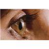
Age-related macular degeneration (AMD) is a disease that affects the macula, the part of the eye that allows a person to see fine detail. AMD gradually destroys the sharp, central vision, which is needed for seeing objects clearly, and for common daily tasks such as reading and driving.

AMD causes no pain and, in some cases, advances so slowly that people notice little change in their vision. In others, the disease progresses faster and may lead to a rapid loss of vision in both eyes. AMD is a leading cause of vision loss in the western world for individuals over the age of 60, and is thought to affect over three million people in the UK alone. AMD occurs in two forms: wet and dry.

Where is the macula?

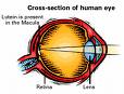
The macula is located in the centre of the retina – the light-sensitive tissue at the back of the eye. The retina instantly converts light into electrical impulses, known as nerve signals, and sends them on to the brain for image interpretation.

What is dry AMD?

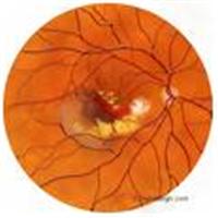
Dry AMD occurs when the light-sensitive cells in the macula slowly break down, gradually blurring central vision in the affected eye. As dry AMD gets worse, a person may see a blurred spot in the centre of his or her vision. Over time, central vision is gradually lost in the affected eye.

The most common symptom of dry AMD is slightly blurred vision. A person may have difficulty recognising faces and may need more light for reading and other tasks. Dry AMD generally affects both eyes, but vision can be lost in one eye while the other eye seems unaffected. One of the most common early signs of dry AMD is ‘drusen’ in the eye, possibly combined with pigment abnormalities.

What is drusen?

Drusen are yellow deposits under the retina often found in people over age 60, and in isolation do not normally cause vision loss. In fact, scientists are unclear about the connection between drusen and AMD, but have found that an increase in the size or number of drusen raises a person’s risk of developing either advanced dry AMD or wet AMD.

An eye care professional can detect drusen during a comprehensive dilated eye exam.

What is wet AMD?

Wet AMD is another advanced stage of AMD and is generally preceded by early or intermediate dry AMD. Wet AMD occurs when abnormal blood vessels behind the retina start to grow under the macula. These new blood vessels tend to be very fragile and often leak blood and fluid. The blood and fluid raise the macula from its normal place at the back of the eye, and damage to the macula occurs rapidly.

With wet AMD, loss of central vision can occur quickly.

What are the stages of AMD?

AMD has three stages, all of which may occur in one or both eyes:

1. Early AMD – people with early AMD have either several small, or a few medium-sized drusen. At this stage, there are no other symptoms and no vision loss.

2. Intermediate AMD – people with intermediate AMD have either many medium-sized or one or more large drusen. Some people see a blurred spot in the centre of their vision. More light may be needed for reading and other tasks.

3. Advanced AMD – in addition to drusen, people with advanced dry AMD or advanced wet AMD have a breakdown of light-sensitive cells and supporting tissue in the central retinal area. This breakdown can cause a blurred spot in the centre of the vision. Over time, the blurred spot may get bigger and darker, taking more of the central vision. Individuals may have difficulty reading or recognising faces until they are very close.

If a person has vision loss from dry AMD in one eye only, he or she may not notice any changes in overall vision. With the other eye seeing clearly, the individual can still drive, read and see fine details, and may notice changes in vision only if AMD affects both eyes.

Which is more common – the dry form or the wet form?

The dry form is much more common, although scientists are still not sure what causes it. More than 85 per cent of all people with intermediate and advanced AMD combined have the dry form.

However, if only advanced AMD is considered, about two-thirds of patients have the wet form. Because almost all vision loss comes from advanced AMD, the wet form leads to significantly more vision loss than the dry form.

Can the dry form turn into the wet form?

Yes. All people who have the wet form of AMD had the dry form first. Dry AMD can advance and cause vision loss without turning into the wet form of the disease. The dry form also can suddenly turn into the wet form, even during early stage AMD. There is no way to tell if or when dry will turn into wet AMD.

The dry form has early and intermediate stages. Does the wet form have similar stages?
No. The wet form is considered advanced AMD.

Can advanced AMD be either the dry form or the wet form?

Yes. Both the wet form and the advanced dry form are considered advanced AMD, and vision loss can occur with either form, although in most cases, only advanced AMD can cause vision loss. People who have advanced AMD in one eye are at especially high risk of developing advanced AMD in the other eye.

Causes and Risk Factors

Who is at risk of developing AMD?

The greatest risk factor is age. Although AMD may occur during middle age, studies show that people over age 60 are clearly at greater risk than other age groups. For instance, a large study found that middle-aged people have about a two per cent risk of developing AMD, but this risk increased to nearly 30 per cent in those over age 75.

Other risk factors include:

• Smoking – smokers have an increased risk of developing AMD

• Family History – individuals with immediate family members who have AMD are at a higher risk of developing the disease

• Obesity – research studies suggest a link between obesity and the progression of early and intermediate stage AMD to advanced AMD

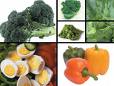
• Poor nutrition lacking in lutein /zeaxanthin rich diet (nutrients found in green leafy vegetables such as curly kale and egg yolk)

• Race – Caucasians seem to be more likely to lose vision from AMD than those with darker skin

• Gender – women appear to be at greater risk than men

Can a person’s lifestyle make a difference?

A person’s lifestyle can play a role in reducing the risk of developing AMD. So it’s important to remember to:

• Eat a healthy diet that’s high in fruit, fish and vegetables, specifically green leafy vegetables

• Avoid smoking

• Maintain a normal blood pressure

• Maintain a healthy weight and exercise regularly

Symptoms and Detection

What are the symptoms of AMD?

Dry AMD: The most common early sign for dry AMD is blurred vision. As fewer cells in the macula are able to function, people will see details such as faces or words in a book less clearly.

Often this blurred vision disappears in brighter light. If the loss of these light-sensing cells becomes great, people may see a small but growing blind spot in the middle of their field of vision.

Wet AMD: The classic early symptom for wet AMD is that straight lines appear crooked. This results when fluid from the leaking blood vessels gathers and lifts the macula, distorting vision. A small blind spot may also appear in wet AMD, resulting in loss of one’s central vision.

Neither dry nor wet AMD cause pain.

How is AMD detected?

An eye care professional may suspect AMD if the person is over age 60 and has had recent changes in central vision. AMD is detected during a comprehensive eye exam that includes a visual acuity test, dilated eye exam, and tonometry (a measurement of intraocular pressure).

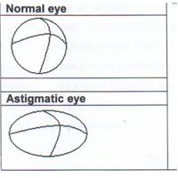
An eye care professional also may perform other tests to learn more about the structure and health of the eye. For example during an eye exam, the patient may be asked to look at an Amsler grid – a grid of straight lines with a black dot in the centre. The patient will be asked to cover one eye and stare at the black dot. While staring at the dot, they may notice that the straight lines in the pattern appear wavy, and that some of the lines are missing. These may be signs of AMD.

If an eye care professional believes the patient needs treatment for wet AMD, he or she will suggest a ‘fluorescein’ and/or ‘indocyanin green angiogram’ to identify any leaking blood vessels and recommend treatment.

What does AMD mean for the patient?

For many, AMD is a shock which can be compounded by lack of information, empathy and support available. Patients are frequently told that little can be done to treat the condition, leaving them feeling both angry and depressed.

People with AMD are more likely to become depressed than the general population, and depression can increase the difficulty of adjusting to the disease (Quality of life in age-related macular degeneration – Royal Holloway University of London, 2006). Indeed, a US cross-sectional study of 151 patients living with AMD reported that the rate of depressive disorder was twice that generally found among elderly people living in the community.

Those living with AMD may lose their independence, requiring help with personal and household tasks and other aspects of daily life. Shopping, cooking and general mobility are also more difficult for people with AMD than those of a similar age with no visual impairment. An additional prospective study of AMD patients with recent (within six weeks) loss of vision to their second eye found that of the 51 participants, 33% met the criteria for clinical depression (a higher rate than 16% found in the wider community), of whom only one was receiving treatment for depression, suggesting low levels of pre-existing depression (Source: Rovner, Casten and Tasman – Effect of depression on vision function in age-related macular degeneration (Journals of Ophthalmology, 2002).

Other health problems associated with age such as arthritis and osteoporosis serve to impair quality of life still further.

Visual hallucinations, known as Charles Bonnet Syndrome, and common in those with visual impairments, can also occur with AMD. While benign and frequently short-lived, research suggests that few are warned that AMD can cause hallucinations and may not report them, featuring dementia. Such unnecessary worry may further damage quality of life.

Rehabilitation, including the provision of low vision aids and training in their use, has been proven to benefit those with AMD, improving visual function and assisting quality of life. Psycho-social interventions, such as peer support groups, also help sufferers adjust to the disease.

Treatment

How is wet AMD treated?

Wet AMD can be treated with laser surgery, photodynamic therapy, and injections into the eye. None of these treatments are a cure for wet AMD, and the disease and loss of vision may progress despite treatment.

1. Laser surgery – this procedure, which is performed in a doctor’s office or eye clinic, uses a laser to destroy the fragile, leaky blood vessels. A high-energy beam of light is aimed directly onto the new blood vessels and destroys them, preventing further loss of vision.

However, laser treatment may also destroy some surrounding healthy tissue and some vision. Only a small percentage of people with wet AMD can be treated with laser surgery. It is more effective if the leaky blood vessels have developed away from the fovea, the central part of the macula.

The risk of new blood vessels developing after laser treatment is high. Repeated treatments may be necessary, and in some cases, vision loss may progress despite repeated treatments.

2. Photodynamic therapy – a drug called ‘verteporfin’ is injected into the arm. It travels through the body, including the new blood vessels in the eye, and tends to “stick” to the surface of the new vessels.

Next, a light which activates the drug is beamed into the eye for around 90 seconds, which destroys the new blood vessels and leads to a slower rate of vision decline. Unlike laser surgery, this drug does not destroy surrounding healthy tissue. Because the drug is activated by light, the patient must avoid exposing skin or eyes to direct sunlight or bright indoor light for five days after treatment.

Photodynamic therapy slows the rate of vision loss, but does not stop it or restore vision in eyes already damaged by advanced AMD. Treatment results often are temporary and a person may need to be treated again. Photodynamic therapy is relatively painless, takes about 20 minutes and is normally performed in a dedicated hospital clinic.

3. Injections – wet AMD can now be treated with new drugs that are injected into the eye (anti-VEGF or anti-angiogenic therapy). Abnormally high levels of a specific growth factor occur in eyes with wet AMD and promote the growth of abnormal new blood vessels. This drug treatment blocks the effects of the growth factor.

A person will need multiple injections that may be given as often as every month. The eye is numbed before each injection, and the patient may need to remain in hospital for a period of time after each session for monitoring. This drug treatment can help slow down vision loss from AMD and in some cases improve sight.

How is dry AMD treated?

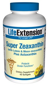
Once dry AMD reaches the advanced stage, no form of treatment can prevent vision loss. However, intake of certain antioxidant vitamins and zinc can delay and possibly prevent intermediate AMD from progressing to the advanced stage, in which vision loss occurs.

The US National Eye Institute’s Age-Related Eye Disease Study (AREDS) found that taking Bausch & Lomb’s PreserVision, a high-potency nutritional supplement containing antioxidants and zinc, significantly reduces the risk of advanced AMD and its associated vision loss. Slowing AMD’s progression from the intermediate stage to the advanced stage will save the vision of many people.

Age-Related Eye Disease Study (AREDS)

What is the dosage of the AREDS formulation?

The specific quantities of antioxidants and zinc used by the AREDS researchers were 500 milligrams of vitamin C; 400 International Units of vitamin E; 15 milligrams of beta-carotene (often labelled as equivalent to 25,000 International Units of vitamin A); 80 milligrams of zinc as zinc oxide; and two milligrams of copper as cupric oxide. Copper was added to the AREDS formulation containing zinc to prevent copper deficiency anaemia, a condition associated with high levels of zinc intake.

Bausch & Lomb’s PreserVision Original formulation, which was proven safe and effective by the AREDS study, is available in soft gel and tablet forms. The new PreserVision Lutein soft gels replace beta-carotene with lutein and are suitable for smokers.

Who should take the AREDS formulation?

People who are at high risk for developing advanced AMD should consider taking the formulation in consultations with their health care provider or personal physician. A person is at high risk of developing advanced AMD if he or she has either:

1. Intermediate AMD in one or both eyes.
-OR-
2. Advanced AMD (dry or wet) in one eye, but not the other.

The AREDS formulation is not a cure for AMD, nor will it restore vision already lost from the disease. However, it may delay the onset of advanced AMD. It may also help people who are at high risk of developing advanced AMD keep their vision.

Can diet alone provide the same high levels of antioxidants and zinc as the AREDS formulation?

No. High levels of vitamins and minerals are difficult to achieve from diet alone. However, previous studies have suggested that people who have diets rich in fruit, fish and vegetables, specifically, green leafy vegetables, have a lower risk of developing AMD.

Can a daily multi-vitamin alone provide the same high levels of antioxidants and zinc as the AREDS formulation?

No. The formulation’s levels of antioxidants and zinc are considerably higher than the amounts in any daily multi-vitamin. If a person is already taking daily multi-vitamins and his or her doctor suggests taking the high-dose AREDS formulation, it is recommended that the individual review all vitamin supplements with his or her doctor prior to taking the AREDS formulation.

Because multi-vitamins contain many important vitamins not found in the AREDS formulation, a person may wish to take a multi-vitamin along with the AREDS formulation. For example, people with osteoporosis need to be particularly concerned about taking vitamin D, which is not in the AREDS formulation.

How can a person take care of his or her vision once diagnosed with AMD?
If a person has dry AMD, he or she should have a comprehensive dilated eye exam at least once a year. An eye care professional can monitor his or her condition and check for other eye diseases. Also, if a person has intermediate AMD in one or both eyes, or advanced AMD in one eye only, the doctor may suggest that the individual take the AREDS formulation containing the high levels of antioxidants and zinc.

Because dry AMD can turn into wet AMD at any time, a person should obtain an Amsler grid from their eye care professional. It is recommended that the individual use the grid every day to evaluate his or her vision for signs of wet AMD. This quick test works best for people who still have good central vision. If a person detects any changes in the appearance of this grid or in his or her everyday vision while reading the newspaper or watching television, he or she needs a comprehensive dilated eye exam.

If a person has wet AMD, a doctor would normally advise immediate treatment. After laser surgery or photodynamic therapy or anti-VEGF anti-angiogenic therapy, a person will need frequent eye exams to detect any recurrence of leaking blood vessels.

Studies show that people who smoke have a greater risk of recurrence than those who don’t. In addition, a person should check his or her vision daily at home using the Amsler grid and will need to schedule an eye exam immediately if any changes are detected.

What can a person do if he or she has already lost some vision from AMD?
If a person has lost some sight from AMD, he or she should not be afraid to use his or her eyes for reading, watching TV, and other routine activities. Normal eye use will not cause further vision damage. These individuals should also ask their eye care professional about low-vision services and devices that may help make the most of their remaining vision.

Many community organisations and agencies offer information about low-vision counselling, training, and other special services for people with visual impairments. Macular Disease Society is of particular value to many existing AMD sufferers. The Royal National Institute of the Blind (RNIB) also provides support and services for those experiencing vision difficulties and loss.

Current Research

What AMD research is currently being conducted?
Research is conducted globally to help provide better ways to detect, treat, and prevent vision loss through AMD. Currently, scientists are:

• Studying the possibility of transplanting healthy cells into a diseased retina – a London-based project to cure AMD has recently been launched, following £4 million donation from an anonymous American donor. This will involve a study where patients will be treated for dry AMD with injections of Retinal Pigment Epithelium (RPE) into the eye. The RPE itself has been developed under laboratory conditions from embryonic stem cells. The study is likely to take at least five years to complete, and will be undertaken by Moorfields Eye Hospital in conjunction with the Institute of Ophthalmology at University College London and the University of Sheffield

• Evaluating families with a history of AMD to understand genetic and hereditary factors that may cause the disease

• Looking at certain anti-inflammatory treatments for the wet form of AMD
• AREDS II further examining the role of vitamins, lutein, omega-3 fatty acids and zinc

image

Lutein & Zeaxanthin diet supplements slow down ageing eye disease

image
image

Belfast: Age-related macular degeneration (AMD), a disease of the eye which is the leading cause of blindness for adults aged over 60, may be accelerated by deficiencies in the diet.

And new research has confirmed that two nutrients in particularl, lutein and zeaxanthin can prevent and even slow down the disease. AMD attacks the macular, which is responsible for sharp central vision and is a disease over and above the normal wear and tear experienced by ageing eyes.

But Professor Usha Chakravarthy of Queen’s University Centre of Vision and Vascular Science and Dr Stephen Beatty of the Waterford Institute of Technology reported that a supplement containing high amounts of the carotenoids lutein and zeaxanthin in addition to the antioxidant nutrients zinc and vitamins C and E, helped preserve macular pigments in patients with age related macular degeneration (AMD), retarding the progression of early to late stage disease. Macular degeneration is the leading cause of vision loss among older individuals residing in western nations.

Professor Chakravarthy and colleagues administered the nutritional supplement or a placebo to 433 participants with early macular degeneration enrolled in centers in Belfast and Waterford, Ireland. The subjects, whose average age was 77 upon enrollment, were followed from October, 2004 to March, 2008.

While participants who received the placebo experienced a steady decline in protective macular pigments, these pigments were preserved in those who received lutein and zeaxanthin.

“Late AMD causes severe sight loss and has a huge economic impact both in terms of the effects of sight loss itself and in terms of the expensive treatments that are needed to deal with the condition,” Dr Chakravarthy stated.

“Up to 500 people a year in Northern Ireland will lose sight in one or both eyes as a result of late AMD . . . Prevention of progression to late AMD can result in a reduced financial and societal burden.”

“These findings are important because this is the first randomized controlled clinical trial to document a beneficial effect through improved function and maintained macular pigments,” he noted. “Further research is needed to confirm these findings.”

There are two types of AMD – dry when the light sensitive cells in the macular break down and wet when the normal blood vessels in the retina start to grow under the macular.

Lutein and Zeaxanthin are thought to be particularly helpful to eye health when combined with a mult-vitamin.

Read more about eye health Eye Care

Omega 3 fatty acids keep eyes younger

image

London: Increased intake of omega-3 fatty acids found in oily fish and fish oil, could help to reduce progression of a serious eye disease that causes blindness in older people, reveals research published ahead of print in the British Journal of Ophthalmology.

Commenting on the study, Dr Ruxton, an independent advisor to the Health Supplements Information Service, notes: “The researchers based their findings on nearly 3,000 people, all of whom were taking part in a supplements trial called the Age Related Eye Disease Study. Participants were randomly allocated to receive either a daily placebo pill or one of three active pills namely: (1) antioxidant vitamins C (500 mg), E (268 mg) plus beta carotene (15 mg); or (2) minerals zinc (80 mg) and copper (2 mg); or (3) antioxidants plus zinc.

“Participants were also quizzed about their diets, using a validated food frequency questionnaire, and were periodically given general physical and eye exams. Detailed photographs of the macula (area at the back of the eye) were taken at the start of the study, after two years, and then again annually until eight years. The trial participants were monitored during this time for the development of age-related macular degeneration (ARMD).

“An earlier publication showed that the antioxidants plus zinc supplement significantly reduced the risk of ARMD. The current study showed that people in the placebo group had a 25% lower risk of developing ARMD if their intake of omega 3 fatty acids from fish was high.

“These findings agree with two other studies from 2009 which also found that omega-three fatty acids were linked with reduced risk of ARMD. An Australian meta-analysis also concluded that consumption of omega 3 fatty acids appeared to be protective against ARMD development.

“Consumption of omega 3 fatty acids in the UK is significantly lower than recommendations. The Food Standards Agency suggests that adults consume two portions of fish a week, one of which should be oily. This equates to 450mg omega 3 fatty acids per day. Among people in Britain who do eat oily fish – less than a third of the population – the average daily intake of omega 3 fatty acids is only 270mg. Amongst those who never eat oily fish, the average daily omega 3 intake is a dismal 147mg . In the context of these low intakes and the increasing evidence that omega 3s seem to lower the risk of ARMD, a supplement containing omega 3 fatty acids could make a useful contribution to eye care.”

Note: ARMD is a progressive degenerative condition, which affects the back of the eye (macula). It robs people of the capacity to see fine detail, and eventually blinds them. In the UK, an estimated 200,000 people have visual loss due to AMD. This number will increase as the population ages. It is estimated there will be 239,000 people visually impaired due to AMD in the UK by 2011. Over half of 33,000 people certified as blind or partially sighted in 1999/2000 were affected by AMD.

[1] Chiu C-J, Klein R, Milton RC, Gensler G, Taylor A. Does eating particular diets alter the risk of age related macular degeneration in users of Age-Related Eye Disease Study Supplements? Br J Ophthalmol 2009, doi: 10.1136/bjo.2008.143412

[2] Chong EW et al. Dietary omega-3 fatty acid and fish intake in the primary prevention of age-related macular degeneration: a systematic review and meta-analysis. Arch Ophthalmol. 2008 Jun;126(6):826-33

[3] Mason P. Towards a healthier Britain. The Potential Role of Food Supplements in Government Food Policy. Available from HSIS.

[4] Bunce C, Wormald R. Leading causes of certification for blindness and partial sight in England & Wales. BMC Public Health 2006;6(1):58

[5] Owen CG, Fletcher AE, Donoghue M, Rudnicka AR. How big is the burden of visual loss caused by age-related macular degeneration in the UK? Br.J.Ophthalmol. 2003;87:312-7

[6] Bunce C, Wormald R. Leading causes of certification for blindness and partial sight in England & Wales. BMC Public Health 2006;6(1):58

Thousands have glaucoma without knowing, say experts

image

London: Glaucoma affects up to half a million people in the UK and is the second largest cause of unnecessary sight loss.[1]

Around a quarter of a million people don’t know they have the condition as glaucoma often doesn’t have any symptoms in its early stages.[1] Boots Opticians urges you to have regular sight tests, which are crucial to help detect early signs of the disease.

Don’t wait until it’s too late. To book an eye test at Boots Opticians, call 0845 120 4343 or visit www.bootsopticians.com

Carolyn Zweig, Boots Opticians says: ‘Regular eye tests are a vital health check, which not only test your sight, but they can also detect eye conditions such as glaucoma and age-related macular degeneration, before you notice the effect on your sight. How often you have an eye test depends on your age and the recommendation of your optometrist, but as a general rule, children should be taken for an eye examination by the age of five and eye tests should be taken every two years after that until the age of 70. At 70 and beyond, you should have an eye test every year.’

Facts about glaucoma

The first World Glaucoma Day was March 6th 2008
There are four types of glaucoma – the most common is open angle or chronic glaucoma

Chronic glaucoma causes gradual blockage of the eye’s drainage tubes leading to a build-up of pressure which damages the optic nerves
50 per cent of glaucoma sufferers go undiagnosed in developed countries[2]
Up to 95 per cent of glaucoma sufferers go undiagnosed in the developing world [2]
Around two in every 100 people over 40 in the UK have glaucoma [3] which equates to approximately 600,000 people

[1] RNIB.org.uk

[2] World Glaucoma Association

3 NHS
4 Based on figures from The Office of National Statistics
5 The College Of Optometrists

Eurocanarias Oftalmológica (eye health), Las Palmas Gran Canaria

image
image

Eurocanarias Oftalmológica
Calle León y Castillo 211
Las Palmas de Gran Canaria
T: + 34 928 49 10 90
www.eurocanariasoftalmologica.com

15% discount

Eurocanarias Oftalmológica is a company devoted to eye surgery and ophthalmic treatments, and has been at the cutting edge of ophthalmology for almost 20 years. Facilities here are state-of-the-art.

Eurocanarias Oftalmológica provides a complete ophthalmology service that covers all of the areas of this speciality, thereby offering on Las Palmas de Gran Canaria a service that matches that of any other leading international ophthalmology centre.

Services

The company offers a full range of eye procedures and treatments makes use of the latest technology and equipment to diagnose and treat eye problems as well the services of ten prominent ophthalmologists

Eurocanarias Oftalmológica is a company devoted to Eye Medicine and Surgery. This clinic has been on the cutting edge of national ophthalmology for almost 20 years.

Eurocanarias Oftalmológica provides a complete ophthalmology service that covers all of the areas of this speciality, thereby offering on Las Palmas de Gran Canaria a service that matches that of any other leading international ophthalmology centre. To do so, the company makes use of the latest technology to diagnose and treat eye problems as well as a provider directory that includes ten prominent ophthalmologists.

Treatments offered include a range of the latest medical interventions for eye health including laser surgery from Myopia, Hyperopia, Astigmatism and other surgical interventions for cataracts and glaucoma as well as contact lenses.

Stem cells used to grow frog eye

image

New york: In an experiment at the SUNY Upstate Medical University, in Syracuse, NY researchers have grown frog eyes from stem cells.

Researcher Michael Zuber and his colleagues took the cells from frog eggs.Then got them to become eyes, by genetically modifying them, inserting transcription factors (proteins that trigger expression of other genes) which are known to regulate eye growth and development.

The scientists then implanted the cells into tadpoles missing an eye. The cells properly developed and differentiated into all seven types of retinal cells and appeared to have the proper structure. Additionally the new eye attached properly to the brain. In swimming tests the eye was shown to be working as implanted tadpoles only swam to the white side of the tank (normal behavior), while blind ones would also swim to the black side of the tank.

Would the technique work on mammals? The answer is maybe — frogs naturally have a much easier type regrowing tissues than humans, in fact they can be triggered to regrow legs and many amphibians can regrow lost tails. Triggering proper differentiation in mammals is much more complex.

Nonetheless, Professor Zuber hopes that chemicals will be found from the research that can activate transcription factors in humans. Even if a full eye could not be grown, this could help people with retinal disorders regenerate ocular tissue.

In a separate, but perhaps equally intriguing study performed by Sujeong Jang of Chonnam National University, in South Korea, and his colleagues, the researchers were able to restore the hearing of deaf guinea pigs by implanting them with human neural stem cells obtained from human bone marrow.

Fat causes eye bags, say scientists

Los Angeles: Scientists have come up with the cause of bags underneath the eyes. And the answer is fat.

All is revealed in the September issue of the Journal of Plastic and Reconstructive Surgery.

Reserachers at the University of California Los Angeles used high-resolution magnetic resonance imaging (MRI) to discover that the amount of fat under the eye increases with age.

Lead author Dr Sean Darcy said that cosmetic surgeons commonly believed that it was a stretching of the skin under the eye which caused the problem.

“However, our study showed there is actually an increase in fat with age, and it is more likely that the fat increase causes the baggy eyelids rather than a weakened ligament,” Darcy said. “There have been no studies to show that the orbital septum weakens.”

The study examined 40 subjects (17 males and 23 females) between the ages of 12 and 80. The findings showed that the lower eyelid tissue increased with age and that the largest contributor to this size increase was fat increase.

According to a recent report by the American Society of Plastic Surgeons, nearly 241,000 Americans underwent eyelid surgery in 2007, making it one of the top four surgical cosmetic procedures performed.

Currently, many plastic surgeons performing procedures to treat baggy eyelids do not remove any fat at all. They reposition the fat or conduct more invasive tightening of the muscle that surrounds the eye, or they tighten the actual ligament that holds the eyeball in place. These procedures are performed despite there being no data indicating that these structures change with age.

“Our findings may change the way some plastic surgeons treat baggy eyes,” said study co-author Dr. Timothy Miller, professor and chief of plastic surgery at the Geffen School. “Our study showed that a component of a patient’s blepharoplasty procedure should almost routinely involve fat excision rather than these procedures.”

Blepharoplasty refers to surgical rejuvenation of the upper or lower eyelids, or both, depending on the extent of aging or disease. The procedure is usually performed on the lower eyelid because the most common complaint patients have is that their eyes appear tired, puffy or baggy. The surgeon makes external incisions along the natural skin lines of the eyelid to remove the excess fat and improve the contour of the lower eyelid.

“Although baggy lower eyelids are a significant result of aging and fat expansion, there are other factors that can contribute too,” Miller said. “We recommend that surgeons evaluate each component and address them accordingly in an individualized approach to blepharoplasty.”

The next phase of research will be to perform MRIs of people with baggy eyelids.

The study was supported in part by a UCLA research-enabling grant and a U.S. Public Health Service grant.

Ageing eyes may get protection from antioxidants

image

London: Eating foods rich in antioxidants may protect ageing eyes from degeneration, according to new research.

Scientists have found a link between two processes in the retina that, in combination, contribute to a disease called macular degeneration.

They found antioxidants disrupt the link and extend the lifetime of irreplaceable photoreceptors and other retinal cells.

“The implication is that people at risk of macular degeneration could help prevent the disease by consuming antioxidants,” said Heidi Vollmer-Snarr, a Brigham Young University chemist who earned a doctorate from Oxford and began work on this disease as a postdoctoral researcher at Columbia.

People struck with the disease first lose central vision and temporarily adjust by relying more on peripheral vision. Some eventually lose their vision entirely.

The study found a destructive synergy between the build-up of a compound called A2E and damage to cellular ‘power plants’ called mitochondria. A2E is a natural byproduct of cellular activity that, unlike other compounds, won’t break down or be disposed by the body.

A problem occurs when A2E encounters oxidative stress created by light exposure. In these circumstances, A2E disrupts energy production in mitochondria.

The resulting energy shortage pulls the plug on daily cleaning and maintenance of photoreceptors and another type of retinal cell.

The result is more A2E buildup, and the cycle of destruction hastens the death of these vital visual cells, which are not replaced when they die.

The experiments performed with visual cells from rats, cows and humans showed that antioxidants could completely counter the damage.

Seeing this process play out in the retina has given Vollmer-Snarr a novel idea for attacking harmful growths in the body.

The strategy would involve delivering potentially disruptive compounds like A2E to the target and then using light to trigger the damage.

The study appears online and is published in an issue of the Journal of Biological Chemistry.

Spire offers new injection for age-related blindness

image

London: A new quick procedure not widely available on the NHS which treats age related blindness disease is now available at 13 Spire Healthcare hospitals around the UK.

Age-related macular degeneration (AMD) affects the central part of your vision and occurs mainly in older people. About one in 100 people aged 65-75 and one in eight people aged over 85 suffer from serious AMD that can result in complete loss of central vision.

There are two types of AMD known as the wet kind and the dry kind. Wet AMD is more destructive and is characterised by abnormal blood vessels growing at the back of the eye. These extra blood vessels leak and can cause rapid loss of sight.

Mr Timothy Dabbs, a specialist eye consultant at Spire Leeds Hospital said:
“Although Wet AMD progresses rapidly and is destructive it is treatable. We inject a special drug under a local anaesthetic to control the growth of abnormal blood vessels in the eye.

“On average a patient will need six injections, they are quick procedures
and patients can normally go home about an hour later. Although they may
not regain full sight, the treatment will often prevent further damage and in some instances can reverse some of the damage already caused.”

Erika Bennett, a patient recently treated at Spire Leeds Hospital, said:
“From not being able to read at all I can now read with my reading glasses.
There is a small improvement with every injection and the treatment itself isn’t painful.

“The staff at the hospital made me feel very comfortable. I had lots of confidence and trust in Mr Dabbs as he made sure he explained every detail of the procedure to me. I would recommend anyone suffering with this condition to try this treatment.”

How to tell if you have AMD
If you’re concerned that you or someone you know may be suffering from AMD, the best self test is to look directly at a straight line such as a door or window frame and using only one eye at a time to see if there is a noticeable kink in the frame which appears to move with your gaze. Your optician will be able to confirm if there is deterioration in your eyes.

The Hospitals currently offering this treatment include:

Spire Bushey Hospital

Spire Cambridge Lea Hospital

Spire Cardiff Hospital

Spire Edinburgh Hospital

Spire Gatwick Park Hospital

Spire Harpenden Hospital

Spire Leeds Hospital

Spire Leicester Hospital

Spire Manchester Hospital

Spire Norwich Hospital

Spire Southampton Hospital

Spire Sussex Hospital

Spire Wellesley Hospital

About Spire
Spire Healthcare is one of the leading independent hospital providers in the UK, with a 25 year heritage of customer service and clinical excellence. It was formed in 2007 from the sale of BUPA Hospitals to leading private equity company Cinven.

Spire Healthcare has 25 hospitals across the UK, providing services for private and insured patients as well as NHS funded patients under the government’s Choose and Book scheme. It also offers cosmetic and weight-loss surgery.

Its hospitals carry out more than 160,000 in-patient and day-case treatments a year and work with over 3,000 consultants. It is proud to be the first independent hospital group to publish clinical outcome data.

Smokers most at risk of eyesight loss from ageing

image

New York: Smoking is one of the strongest risk factors for developing age-related macular degeneration (AMD). But smokers may not benefit from antioxidant vitamin supplements, say experts.

Emily Chew, MD, Deputy Director, Division of Epidemiology and Clinical Research, National Eye Institute, Bethesda, Maryland, United States, said patients who smoke and take beta carotene supplements have an increased risk of developing lung cancer.

In a presentation to the American Academy of Ophthalmology (AAO), Dr Chew outlined which patients should and should not take supplements. In a recent study more than 4,700 patients who received supplements of vitamin C and E, beta carotene, zinc and copper and were followed for 6.3 years.

The results of the study showed that while there was a protective benefit for patients with large bilateral macular drusen and those with advanced AMD in one eye, there was no protective benefit for patients with mild and moderate AMD. The supplements did not prevent progression to severe AMD in these patients.

Dr Chew pointed out, however, that the risk of developing AMD in these patients is “exceedingly low,” and therefore, it is unlikely that they would progress to severe AMD.

According to Dr Chew, patients who still smoke, or quit smoking within the past year should avoid the supplements because of the increased risk of cancer. Nor should the offspring of patients with AMD take the supplements, unless they too have AMD in one eye, or large bilateral drusen.

But for most individuals, the benefits of the supplements seem to outweigh any risks. Recently, a meta-analysis of 68 trials reported no significant impact associated with a wide variety of antioxidant supplements. More study is needed, Dr Chew said
Dr Chew concluded that the public health impact of this supplementation regimen could prevent 300,000 people over the next 5 years from either developing advanced macular degeneration or experiencing significant loss of vision as a result of progressive disease.

FDA seizes $2m of Jan Marini eyelash rejuvenator over eyesight threat

image

Washington: At the request of the U.S. Food and Drug Administration, U.S. Marshals seized today 12,682 applicator tubes of Age Intervention Eyelash, a product that may, in some users, lead to decreased vision. Authorities said the sales value of the seized tubes is approximately $2 million.

Age Intervention Eyelash is sold and distributed by Jan Marini Skin Research, Inc., of San Jose, Calif.

The FDA considers Age Intervention Eyelash to be an unapproved and misbranded drug because Jan Marini Skin Research has promoted the product to increase eyelash growth. Before a new drug product may be legally marketed, it must be shown to be safe and effective, and approved by FDA. The agency takes seriously its responsibility to protect Americans from unapproved drugs.

FDA also considers the seized Age Intervention Eyelash to be an adulterated cosmetic. The product contains a drug called bimatoprost, an active ingredient in an FDA-approved drug to treat elevated intraocular pressure (elevated pressure inside the eye).

For patients using the prescription drug, using the Age Intervention Eyelash in addition to the drug may increase the risk of optic nerve damage because the extra doses of bimatoprost may decrease the prescription drug’s effectiveness. Damage to the optic nerve may lead to decreased vision and possibly blindness.

In addition, use of Age Intervention Eyelash may cause other adverse effects in certain people due to the bimatoprost, including macular edema (swelling of the retina) and uveitis (inflammation in the eye), which may lead to decreased vision.

The U.S. Attorney’s Office for the Northern District of California filed the complaint requesting the seizure, and coordinated with the FDA. The California Department of Public Health‘s Food and Drug Branch had previously embargoed the seized products at the San Jose facility. Jan Marini Skin Research has notified FDA that the company ceased manufacturing and shipping any Age Intervention Eyelash product containing bimatoprost last year.

The FDA recommends that consumers, dermatologists, and estheticians who may still have Age Intervention Eyelash discontinue using it and discard any remaining product. FDA also recommends that consumers consult their health care provider if they have experienced any adverse events that they suspect are related to the product’s use.

The injection that cures wet macular eye degeneration

image

London: A new treatment for the “wet” version of age related macular degeneration (AMD) in the form of a painless injection into the eye is available for the first time in the UK.

AMD which attacks the macula, located at the back of the eye at the centre of the retina. The macula enables us to see directly what is in front of us and pick out fine detail. It helps us with everything from reading and driving to recognising faces and seeing colour.

In the early stages of AMD central vision may be blurred or distorted, with objects looking an unusual size or shape and straight lines appearing wavy or fuzzy. Those suffering from the condition may see lights, shapes and colours that are not actually there.

Laurence Whitefield, Consultant Ophthalmic Surgeon at Capio Eye in London’s Harley Street explains: “AMD is the most common cause of poor sight among people over the age of 60 in the UK. It is unclear what causes AMD, but it becomes more likely as a person ages as, with time, the delicate cells of the macula become damaged and worn out.”

There are two forms of AMD – a less severe ‘dry’ form, and the rapidly degenerating ‘wet’ AMD which develops quickly as fragile blood vessels grow behind the retina often leak fluid and blood. This results in scarring and rapid sight loss.

There two breakthrough treatments for wet AMD are known by the brand names Avastin or Lucentis and have been shown to stabilise sight in 95% patients suffering from wet AMD and improve sight in one third. Neither of these treatments are generally available through the UK’s state health service, the NHS.

Mr Whitefield says: “In the US ophthalmologists who specialise in retinal disease have been using Avastin(r) for some months to treat patients with wet or neovascular form AMD. Results have shown that the treatment controls the abnormal blood vessels in AMD, increasing vision. Lucentis is a very exciting development because it can be used to treat all types of AMD.

“Thousands of people a year in the UK get wet AMD leading to rapidly sight loss. This treatment provides patients with a way of recovering their sight. Even patients well below the threshold for registration as blind have been shown to benefit from the treatment”.

Both Avastin and Lucentis treatments consist of three injections into the eye that stop the development of leaky blood vessels that trigger AMD.
Further injections are sometimes necessary, depending on a patient’s progress. The procedure is painless and patients do not need to wear eye pads. The treatment works on patients suffering recent deterioration of vision and is not suitable for those who are already blind or who have established scarring of the retina.

The new drugs are known as anti-VEGF drugs and work by targeting VEGF (Vascular endothelial growth factor), a protein that helps the formation of new blood vessels. In the case of AMD these new blood vessels are unstable and tend to leak fluid and blood under the retina. The resulting scarring then causes irreversible sight loss. The anti-VEGF drugs inhibit the growth of new blood vessels, thereby minimising the risk of scarring.

The costs of the procedure are £500 for the initial consultation with the surgeon and the diagnostic tests to determine whether the treatment is appropriate, then £650 for the first treatment injection and £550 for subsequent treatments. Patients will need the first treatment and then usually at least two more. The speed of results vary by patient.

Contact information: Capio Eye is a private hospital based in London’s Harley Street which offers the very best in eye care, refractive and cataract surgery. www.capioeye.co.uk”>

Please contact us for more information or to make an appointment.

Free phone : 0800 – 169 20 20
Telephone : + 44 (0)20 703 410 30
Fax : + 44 (0) 20 703 410 32
E-mail :
capio-eye-enquiries”>

Address : Capio Eye London
114a Harley Street
London W1G 7EL

Antioxidants do not prevent degenerative eye disease

image

Melbourne: A diet rich in antioxidant vitamins and minerals does not seem to prevent the degenerative eye disease known as age related macular published on www.bmj.com today.

Age related macular degeneration is the leading cause of visual loss in older people. It is caused by the progressive break down of light sensitive cells in the macula, located in the centre of the retina at the back of the eye. Sufferers do not go blind, but find it virtually impossible to read, drive, or do tasks requiring fine, sharp, central vision.

Risk increases with age and smokers are thought to be more susceptible.

Antioxidants (such as vitamin C, vitamin E, various types of carotenoids, and zinc) are thought to reduce oxidative damage to the retina. But the evidence to support the role of dietary antioxidants in preventing macular degeneration remains unclear.

So researchers at the Centre for Eye Research Australia, the University of Melbourne analysed the evidence to examine the role of dietary antioxidants or dietary supplements in the primary prevention of age related macular degeneration.

They identified 11 studies (seven prospective studies and three randomised controlled trials) involving 149,203 people. A range of common dietary antioxidants were investigated and all the studies were carried out amongst well nourished Western populations with an average follow-up period of nine years.

Importantly, all the studies adjusted for age and smoking in their analyses.

The antioxidants investigated differed across studies, but when results were pooled they showed that vitamin A, vitamin C, vitamin E, zinc, lutein, zeaxanthin, α- carotene, β-carotene, β-cryptoxanthin and lycopene have little or no effect in the primary prevention of early age-related macular degeneration.

None of the three trials found antioxidant supplements to be protective in the primary prevention of early age related macular degeneration.

Despite some study limitations, the authors conclude that there is insufficient evidence to support the role of dietary antioxidants, including the use of dietary antioxidant supplements, for the primary prevention of early age-related macular degeneration.

Currently, cigarette smoking remains the only widely accepted modifiable risk factor for the primary prevention of early age-related macular degeneration, and patients seeking advice on this condition should be encouraged to quit, they add.

An accompanying editorial by Jennifer Evans at the International Centre for Eye Health supports these findings and says that reducing the prevalence of smoking is probably the most effective method of reducing the population burden of this common cause of visual loss in older people.

Astigmatism

image

Astigmatism is one of the most common eye conditions – most people have it to degree. It is often mistakenly referred to as ‘stigmatism’.

Astigmatism occurs when the cornea or lens are shaped more like a rugby ball – more curved in one direction than the other. And light is not focused on one point on the back of the eye. For instance, vertical lines may be clearer than two horizontal or vice versa.

This means that the image at the back of your eye is not as sharply focused and vision may be blurred. Corneal astigmatism is when the cornea is a distorted shape and lenticular astigmatism occurs when the lens is distorted.

To focus images, the eye has two parts – the cornea and the lens. The cornea is the curved ‘window’ at the front of the eye. When the cornea and the lens inside the eye area are a regular symmetrical shape all incoming light is sent the same way to make a sharply focused image onto one single point on the retina at the back of the eye. The retina is the light-sensitive area at the back of the eye, like the film in a camera. If light is only focused on one part of the eye, vision will be clear.

Most people with astigmatism are born with the condition, although it can develop throughout life. A condition called keratoconus can also change the shape of the cornea with age and this can cause astigmatism. Rarely, astigmatism can be as a result of an injury or eye disease. Usually a person with astigmatism will have other visual problems, such as long or short sightedness.

Those people with a small degree of astigmatism may not experience any problems. Those whose astigmatism is more significant may experience any or all of the following:

* Blurring and distortion of near or far-away objects
* Headaches when trying to focus
* Senstivity to light
* Tired and dry eyes

In some cases, astigmatism is present at birth as the size and shape of the eye is developed in the womb. It may be due to environmental and genetic factors, but the exact cause is not always known. Sometimes an astigmatism can develop after an eye injury, surgery or eye disease. Astigmatism is not caused by factors such as reading in poor light, squinting or watching too much television.

Although astigmatism is extremely common and afffects most people to some degree, many may not need any corrective action. Most people who wear glasses or contact lenses may have some degree of astigmatism. Treatments include corrective spectacle lenses, special contact lenses or surgery.

People with astigmatism cannot see horizontal or vertical lines clearly at the same time. This is what makes objects appear blurry or out of focus.

For those who have astigmatism, an optometrist, ophthalmic surgeon or eye specialist should carry out a full eye examination to assess the full scale of the problem. Options for correction can be discussed in full. Corrective glasses, special contact lenses or surgery are all options. A regular eye test is vital to maintaining healthy eyes. We should each aim to have our eyes tested every two years at least.

The eyes have it – 10 secrets you should know!

London: The UK’s leading body for eye health professionals, the College of Optometrists, reveals ten eye secrets.

1. Wearing someone else’s glasses may damage your eyes.

False: Although you may not be able to see very well with them and may get a headache or double vision, you won’t come to any harm from wearing glasses that are not your prescription (unless you’re driving a motor vehicle).

2. Watching TV too much or too closely will damage your eyes.

False: Watching too much TV or sitting very close to it may make your eyes tired or give you a headache – particularly if you are watching TV in the dark when you are effectively looking at a moving light, like a torch – but won’t cause any serious permanent damage.

3. Masturbation makes you go blind.

False: The only correlation between the two is that semen contains a large amount of zinc and a deficiency in zinc (although nearly impossible to achieve solely by masturbating) will cause a decline in a person’s vision.

4. Cutting your eyelashes off or putting Vaseline on them makes them grow back or appear fuller.

False: Cutting your eyelashes off makes you look like an idiot and your eyelashes do not grow back fuller. Putting Vaseline on them only gives you a greasy pillowcase.

5. Exercising the eye muscles can allow you to ‘throw away your glasses’.

False: People (normally) need specs because of the shape and size of their eye (i.e. their eyes are too big or too small). Exercises won’t help this.

6. Not wearing your glasses will make you depend upon them less.

False: If you don’t wear your glasses you may become more accustomed to the blur and won’t remember how bad it is, and thereby think that your eyes have got better (when they haven’t).

7. By looking at the patterns, colours and other characteristics of the iris you can tell what health problems a person has.

False: There is no scientific proof for this. However, when an optometrist carries out an eye examination they will not only test your sight, but will also check the health of your eyes and look for signs of some general health problems.

8. Eating carrots will improve your eyesight.

Some truth: Carrots are a source of vitamin A, which is important for the eyes. However, before you embark on an all-carrot diet to improve your vision, note that it is more important for eye health to have a good balanced diet that supports your all-round health. Poor nutrition has been implicated in diseases such as age-related macular degeneration (AMD).

9. Using your eyes too much can wear them out.

False: They will last for your whole life if they are healthy (or have conditions that are treatable). The health of your eyes has nothing to do with the number of hours you use them.

10. Holding books up close will damage a child’s eyes.
False: Where or how your child holds a book has no effect on the health of the eyes or the need for glasses. Sometimes children find it more comfortable to read close-up and their very good focusing ability makes it easy for them to do so.

About the College of Optometrists

1. The College of Optometrists is the Professional, Scientific and Examining Body for Optometry in the UK, working for the public benefit. Supporting its Members in all aspects of professional development, the College provides pre-registration training and assessment, continuous professional development opportunities, and advice and guidance on professional conduct and standards, enabling our Members to serve their patients well and contribute to the wellbeing of local communities.
2. Previously known as ophthalmic opticians, optometrists are trained professionals who examine eyes, test sight, give advice on visual problems, and prescribe and dispense spectacles or contact lenses. They also recommend other treatments or visual aids where appropriate. Optometrists are trained to recognise eye diseases, referring such cases as necessary, and can also use or supply various eye drugs.
3. Optometrists study at university for at least three years and participate in a full year of training and supervision, called the pre-registration year, before qualifying. Once qualified, they have the opportunity to develop their interests in specialist aspects of practice such as contact lenses, treating eye diseases, low vision, children’s vision and sports vision.
4. All optometrists practising in the UK must be registered with the General Optical Council, the profession’s regulatory body, and are listed in the Opticians Register. The letters FCOptom or MCOptom after an optometrist’s name means that he or she is a fellow or member of the College of Optometrists.
5. There are currently over 10,000 registered optometrists in the UK.

Thousands may go blind as UK government denies key drug to elderly

London: The UK government’s drug watchdog is refusing to allow doctors to precribe a drug which could save the sight of thousands of elderly people who live in England and Wales.

At the same time the drug – Macugen – is available on the public health service, the NHS, to those living in Scotland.

Macugen is used to treat the wet form of age-related macular degeneration, where veins leak causing blindness.

The watchdog, the National Institute for Health and Clinical Excellence. said another drug, Lucentis, could be used but only if patients have gone almost blind in one eye and the disease is far progressed in the other.

It also restricted the use of Lucentis to a specific type of the condition which affects only around 20 per cent of sufferers.

There are around 26,000 new cases of wet AMD, which affects the macular region at the back of the eye, in the UK every year.

Based on Nice’s recommendations, 80 per cent of these patients – more than 20,000 a year – will be left to go blind. Macugen and Lucentis, known as antiVEGF drugs, stop the deterioration of the sight.

On Monday the Scottish Medicines Consortium, the rationing body north of the border, approved Lucentis for all NHS patients after previously giving the green light to Macugen.

It is the latest example of post-devolution Scotland giving its patients access to many drugs denied those in England and Wales, which is covered by NICE.

The drugs are also available in many other countries including Germany, France, the Netherlands, Italy, the U.S. and Australia.

Nice’s draft guidance, which is up for consultation, was greeted with anger from charities, clinicians and opposition politicians. The Royal National Institute of Blind People said it was ‘outraged’.

Head of campaigns Steve Winyard said: ‘Anti-VEGF drugs have the potential to halve the number of people going blind each year and patients in the UK who can benefit from them must all have them – and quickly.

‘It is simply unacceptable that Nice is recommending that only a small minority of patients within England and Wales will benefit from these ground-breaking treatments. Nice must reconsider.

‘Anti-VEGF drugs are cost- effective. It is much more expensive to support someone once they have lost their sight than to provide sight-saving treatment.’

Tom Bremridge, chief executive of the Macular Disease Society, said: ‘Limiting the treatment options to 20 per cent of patients who would benefit is unjustifiable and allowing one eye to go blind before treating the second is cruel and totally unacceptable.’

Winifred Amoaku, consultant at the Royal College of Opthalmologists, said the two drugs worked differently. She attacked the fact that only Lucentis had been given the go-ahead because some patients would benefit more from Macugen.

‘Both treatments should be made available to opthalmologists,’ she said. ‘One treatment doesn’t fit all and opthalmologists should be able to prescribe the most suitable option for their patient.’

The Nice ruling comes despite a study published in the New England Journal of Medicine which found that the sight of nine out of ten patients given Lucentis improved or stabilised after two years of treatment.

Lucentis costs up to £2,000 per injection or £28,000 for a full course of treatment, which is 14 injections over two years. Macugen is around £1,800 per injection or £36,000 for a full course of 20 injections.

Dr David Gillen, medical director of Macugen manufacturer Pfizer, said: ‘Macugen has been shown to maintain vision in patients will all types of wet AMD and has a licence to reflect this.

‘From a cost perspective, it has been convincingly demonstrated that Macugen’s cost-effectiveness can be enhanced when treatment is started at an early stage before too much vision is lost.’

Stem cell surgery for ageing eyes available within five years, predict doctors

London: British scientitsts are working on a proecdure to use stem cells as a cure for age-related macular degenertion, a leading cause of blindness in the elderly.

The first 45-minute operation would use stem cells grown in a lab to cure the dry form of the diseease and it is estimated that the procedure will be available within five years.

The improvement in sight is likely to be great enough for the the blind to regain the ability to carry out everyday tasks such as reading or driving.

The pioneering stem cell surgery tackles age-related macular degeneration (AMD), the most common cause of blindness in the elderly. There are about 300,000 sufferers in this country and the number is expected to treble in the next 25 years to around one million as the population ages.

AMD, which affects a quarter of over-60s in the UK and more than half of over-75s to some degree, occurs in two forms. While the ‘wet’ form can be combated with drugs, there is no treatment for the ‘dry’ form which accounts for 90 per cent of cases.

The treatment centres on human embryonic stem cells grown in a laboratory. These are ‘blank’ cells with the power to turn into different cell types and are used to create small patches identical to the cells damaged in the eyes of AMD sufferers.

Packaged into a syringe, the patch is injected into the back of the eye where it replaces damaged cells and restores sight.

The technique is being developed by scientists and doctors from University College London, Moorfields Eye Hospital, also in London, and Sheffield University, working together in the London Project to Cure Blindness.

Their work has been boosted by a £ 4million donation from an anonymous American benefactor.

Project director Professor Pete Coffey said: ‘This could have a tremendous effect on a huge population who have no current therapy.’

The technique has been tested on rats suffering from a condition similar to AMD and their sight was restored.

Further evidence that the technique is likely to succeed comes from human operations. In these, the researchers restored vision using healthy cells taken from the corner of the patient’s own eye.

In some cases, the transplants were so successful that the patients were able to read, cycle and use a computer.

However, such surgery is extremely complex and time-consuming and so unlikely to be suitable for large-scale use. Using ‘readymade’ patches of cells would greatly simplify the operation, making it suitable for use on millions.

The scientists are now working on making such patches, measuring just four by six millimetres, which will be injected into the back of the eye under local anaesthetic in an procedure lasting between 45 minutes and an hour. The patient, who would have to take drugs to stop the cells from being rejected by the body, could go home the same day. After two to three weeks, vision should start to improve.

It is not yet known how long the effects will last but the patients who had transplants of their own cells are still benefiting from the treatment which took place two and a half years ago. While the patches are most likely to benefit those in the early stages of AMD, the researchers believe it should be possible to adapt them to treat those in later stages.

It is hoped that the technique might also benefit those who have lost their sight as a complication of diabetes.

Consultant surgeon Lyndon da Cruz of Moorfields Eye Hospital said that within ten years the procedure could become as commonplace as cataract surgery.

He said: ‘If we can do a single procedure in a person under local anaesthetic in 45 minutes, it’s feasible.

‘The science is something we can work on but the surgery has to be something we can deliver to many people.’

Eye experts said the research offered real hope to sufferers of AMD. Tom Bremridge of the Macular Disease Society said: ‘This development is exciting and encouraging for current and future generations of AMD patients.

‘While treatments for “wet” AMD are advancing rapidly, sadly, patients with “dry” AMD have had no prospect of any viable therapy.’

Professor Alistair Fielder, of the charity Fight for Sight, said the research represented ‘a real chance to tackle an untreatable condition and bring hope to many’.

He added: ‘It is marvellous to think that clinical trials could start within four years.’

Although many believe it is wrong to use embryonic stem cells – plucked from an embryo in the first days of life – in medicine, sophisticated laboratory techniques mean it should be possible to generate a treatment for millions of people from cells derived from a single embryo.

Stem cell research offers hope for treating and curing a host of conditions.

In recent work, British experts have succeeded in growing a ‘ miniliver’ – a tiny bundle of liver cells – from stem cells, while Israeli scientists have grown a tiny section of beating heart tissue from stem cells gleaned from human embryos.

Dirty contact lenses put wearers at risk of blindness

London: Almost three quarters of contact lens wearers are risking blindness because of their dirty habits, according to a new study out today. The new figures show one in five people lick their contact lenses before putting them in their eyes and two out of five apply their lenses with dirty hands.

Shockingly over 15% of wearers will pick an unwashed contact lens off the floor and pop it in their eye and 70% of people keep their lenses in for far too long every day. But careless contact lens wearers rarely realise their bad habits increase the risk of their contracting eye diseases, including conjunctivitis and acanthamoeba keratitis, a rare but very painful and potentially blinding infection of the cornea, the transparent covering at the front of the eye.

Men are the lazy ones when it comes to caring for their eyes, with more than half sleeping in their lenses and 44% not bothering to wash their hands before putting their lenses in. But surprisingly women have the dirtiest habits. Nearly a quarter of women lick their lenses before popping them in their eyes and 16% will use a dirty lens they have dropped on the floor, according to research by the UK’s College of Optometrists. And you have heard of sharing make-up, some women even share their contact lenses with their best friends.

Experts warn even habits that seem harmless such as washing lenses in tap water or with a different cleansing solution can damage the wearers’ eyes. Tap water contains bacteria that can blind you and using the wrong solution not only harms the lens but can also produce allergic reactions or even burn the eye.

Optometrists advise contact lens wearers never to wear other people’s lenses, a lens that is chipped or damaged or put the wrong lens in the wrong eye. The experts add people should always clean their lenses either after wearing them or before putting them in their eyes and to make sure their hands are clean before handling them.
And a final warning, don’t wear your lenses too long or sleep in them and visit an optometrist at least every two years.

Kevin Lewis, President of the College of Optometrists said: “Good eye care is crucial for everyone but even more important for those of us who wear contact lenses. Poor hygiene can lead to infections which range from a simply uncomfortable but temporary problem to a very nasty health issue that may lead to blindness in a matter of days.”

“Our eyes are precious and we need to appreciate that treating them carelessly and with bad hygiene habits is likely to cause problems in the future.”

For more information visit www.college-optometrists.org

British doctors restore eyesight with stem cells

London: British doctors have restored eyesight by using stem cells to replace damaged cells.

Although the results have delighted experts, the charity the Royal National Institute for the Blind warned that sufferers of blindness should not raise their hopes until further trials are carried out.

The treatment could also help sufferers of macular degeneration – the most common cause of blindness in the elderly – and those who have lost their sight as a complication of diabetes.

The researchers, from University College London and London’s Moorfields Eye Hospital, used stem cells – basic cells with the ability to turn into different types of tissue – to restore the vision in blind mice.

The cells were injected into the back of the eye where they replaced damaged photoreceptors – the tiny light-sensitive cells on the retina.

Until now, there was no way of reversing the damage – and previous stem cell transplants had failed.

Central to the success of the technique was the selection of cells which were slightly more mature than those tried by other scientists.

These cells turned into photoreceptors and successfully connected with the nerves leading to the brain. By shining light into the animals’ eyes, the researchers were able to show that vision had been restored to around a quarter of normal levels. Increasing the number and type of cells transplanted could improve sight even further.

The scientists caution that the technique is still in its infancy. But Dr MacLaren, of Moorfields Eye Hospital, said: ‘This research is the first to show photoreceptor transplantation is feasible. We are now confident that this is the avenue to pursue to uncover ways of restoring vision to thousands.’

Exercise may protect eyes from ageing

image

New York: Taking regular exercise may help reduce the risk of age-related eye diseases, scientists at the University of Wisconsin have discovered.

The researchers examined 4,000 men and women over a 15 years period, carrying out eye tests and recording levels of exercise, says their report in the British Journal of Ophthalmology.

Age-related macular degeneration (AMD) destroys the functionality of light sensitive cells behind the eye. The scientists found those with an active lifestyle were 70% less likely to develop the degenerative eye disease than those with a sedentary lifestyle.

AMD s the leading cause of severe vision loss in the over-50s in the developed world and affects central vision, needed for driving. The study of people aged between 43 and 86 began in 1988 and they were assessed every five years.

The research examined their exercise habits and eye health and found one in four had an active lifestyle and nearly one in four climbed more than six flights of stairs a day.

After taking into account other risk factors such as weight, blood fat levels and age, active participants were 70% less likely to develop AMD than those who did little exercise. It also showed regular walkers were 30% less likely to get the disease.

Authors of the report did warn however that diet may also explain the findings.

1 in 4 adults fail to get eye sight check, says new survey from Centrum

London: Our eyesight is our most precious sense1, according to a new survey commissioned by Centrum*2, the world’s number one selling multivitamin, which now contains FloraGLO lutein3, a natural form of lutein that can help maintain healthy eyes.

The research shows that we dread losing our sense of independence the most and not being able to see loved ones if our eyesight was to get worse, yet 1 in 4 of us have never had their eyes checked.

In the UK we value our eyes more than any other sense: 61 per cent say it is the sense that they would least like to lose. Compared to other parts of the body, eyes are also the ones that we are most squeamish about getting injured (63%).

The Centrum survey found that if our eyesight was to get worse, we would be most worried about losing our independence (46%) and not being able to see loved ones (27%), with other concerns being not being able to read (8%), drive safely (6%) or see in an emergency (4%).

However, our priorities appear to shift as we get older, with those over 65 years old being as worried about not being able to see their loved ones as not being able to read (17%). Those living in Wales and East Anglia are particularly loving groups, being the only regions where loved ones (38%/41%) feature higher than personal independence (36%/35%).

The strength of the love for our families is also evident when asked what the most amazing thing is that we have ever seen: nearly half of those surveyed (47%) said it was a family moment, such as the birth of their new baby or grandchild, a christening or a wedding. These overtook spectacular natural sights and landmarks as the most amazing sight. Again, those in Wales are more family-orientated than most, with 62 per cent choosing a family moment as their most amazing sight being pipped only by the 66 per cent in East Anglia.

Keeping our eyes healthy is the main reason that we get our eyes checked (29%), although reminders from opticians (23%) and difficulties with reading (14%) are also important factors. Despite all of this, most people do not appear to be taking the best care they can of their eyes.

One of the things we can do to help keep our eyes healthy is to eat foods that contain lutein, a natural pigment that helps protect our eyes. From research we know that only one in five people in the UK are aware of lutein. 4 Lutein cannot be made by the body, so we have to make sure we get it through the foods we eat. Lutein can be found in spinach, kale, peas, cos or romaine lettuce and broccoli. Although there is no recommended daily allowance of lutein set in the UK, some other countries suggest we need to have about 6mg a day (which equates to about one bowl of spinach a day).

Centrum research showed that less than half of those surveyed (42%) eat foods that contain lutein on a daily basis. Beside food, we can also get lutein through certain vitamin and mineral supplements. Centrum contains vitamins and minerals specifically designed to supplement the daily diet, with Centrum and Centrum Select 50+ now also containing lutein.
-ends-
Notes for editors:
1. The survey, carried out for Centrum by BMRB Omnibus, quizzed 1,000 adults over the age of 16 across England, Scotland and Wales in June 2006.

2. Centrum is a multivitamin that’s complete from A to Zinc. It is a comprehensive formula of vitamins and minerals specifically designed to supplement the daily diet. Three other variants are carefully tailored to the needs of children (Centrum Kidz), the over 50s (Centrum Select 50+) and people who lead an active or hectic lifestyle (Centrum Performance).

3. Lutein is a carotenoid that is found in fruit and vegetables. It has been shown in clinical trials to help protect against retinal damage. FloraGLO lutein is a natural form of lutein that is extracted from marigold plants. It is an ideal form for human health because it’s identical to the lutein found in dark green vegetables. It is easily absorbed by the body, and deposits itself in the eye to absorb potentially harmful blue light and protect the eye.

4. Lutein Brand and Health Awareness Study UK March 2006, produced by Frost & Sullivan on behalf of Kemin.